- Review
- Open access
- Published:
Handling and pathology reporting guidelines for bladder epithelial neoplasms – recommendations from the Brazilian Society of Pathology / Brazilian Society of Urology / Brazilian Society of Clinical Oncology
Surgical and Experimental Pathology volume 7, Article number: 8 (2024)
Abstract
The Brazilian Society of Pathology Guidelines Project aims to provide recommendations for clinicians and pathologists based on the best available scientific evidence. It reviews the currently available and emerging histopathological and molecular aspects of bladder cancer that are necessary for the best patient’s management. This paper is a result of a combined effort of the Brazilian Society of Pathology, the Brazilian Society of Urology, and the Brazilian Society of Clinical Oncology to call attention to the essential pre-analytical issues, the required clinical information and specimen handling to allow proper diagnosis, grading, staging and characterization of the molecular aspects of bladder epithelial neoplasms.
Background
The evolution of personalized medicine requires adopting optimal tissue handling and fixation procedures and adequate clinicopathological correlation. In addition, pathologic examination must produce a pathology report with a precise diagnosis and explicit information with all items of prognostic value that may influence clinical management and treatment. In this combined effort of the Brazilian Society of Pathology, the Brazilian Society of Urology, and the Brazilian Society of Clinical Oncology, we provide recommendations that should guide proper communication by urologists and appropriate examination and reporting by pathologists. The final goal is to improve communication between surgeons, pathologists, clinical oncologists, and radiation oncologists, leading to optimal management of patients with bladder neoplasia.
A template of the reporting guide is available here (see Supplemental file 1), and a Portuguese edition is available on the website of the Brazilian Society of Pathology: https://www.sbp.org.br/guidelines/neoplasias-uroteliais-3/.
Pre-analytical phase
Best laboratory practices
The pathologist is central in communicating with a multidisciplinary team, including nurses, surgeons, oncologists, radiologists, and radiation oncologists. The pathologist must balance the requirements of high-quality samples and rapid results with smaller samples with less invasive procedures (Hansel et al. 2013; Hansel and Lerner 2018). Molecular studies used in clinical practice for managing bladder cancer patients included PD-L1 evaluation by immunohistochemistry, evaluation of FGFR3 aberrations, and large-scale sequencing. As these migrate from the academic centers to the broader setting of surgical pathology (including community laboratories), it became clear that the quality of samples is heterogeneous, resulting in a high rate of samples inadequate for molecular analyses. The type of samples and the procedures associated with their collection and fixation methods are detailed below.
Small biopsies should be immediately fixed in buffered formalin. The grossing team should be cautious to avoid losing small samples: tiny fragments may be sent inside histology cassettes, sealed or on filter paper, to prevent losing samples.
Large surgical specimens (from cystectomy and lymphadenectomy) require further handling in the grossing room before complete fixation. Proper fixation is obtained by reducing the warm ischemia time (from the point that blood supply has been reduced or cut to organ removal) and cold ischemia (time to embed the specimen in fixation media), proper dissection to allow total exposure of all tissues (e.g., bladder mucosa in a large cystectomy specimen) and complete fixation. Cystectomy specimens require opening of the anterior wall (for a total exposure of bladder mucosa to the fixative) or injection of the fixative through the urethra to insufflate the bladder cavity (then clamping the distal urethra). The quality of molecular evaluation thus depends on the quality of gross specimen handling and other factors such as previous therapies, neoadjuvant chemotherapy, radiation therapy, and BCG instillation. A detailed protocol for gross examination of cystectomy specimens is beyond the scope of this report and may be obtained elsewhere (Chandra et al. 2010; Mazzucchelli et al. 2021).
Molecular tests commonly require a previous assessment of the histologic slide to select areas with the highest tumor cellularity and, if possible, avoid large necrotic areas. This evaluation allows the dissection of unstained slides. Thus, it is good practice for the pathologist to select the best block when referring a case to another laboratory to perform the molecular analysis. A hematoxylin and eosin-stained slide should go along with the blocks to avoid the requirement of repeated work and tissue consumption. It is not required to mark specific areas for a molecular test since specific protocols may differ.
The transurethral resection of a bladder tumor (TURBT) allows the obtaining of more significant amounts of fragments than small biopsies. However, thermic / cautery artifacts may be variable and, in extreme cases, may preclude a proper diagnosis. In contrast to the sampling of prostate TURBT (usually associated with benign disease), it is recommended that the entire specimen be submitted for histopathological evaluation, avoiding submitting excessive amounts of tissue in a single cassette, that could lead to impaired processing and overlap in the inclusion procedure.
For all solid samples, the multidisciplinary team should make all efforts to adhere to the best practices suggested by the Brazilian Society of Pathology (Assis 2020):
-
ideal period of 10 min from sample removal to be placed in fixation media (acceptable to last until 60 min).
-
use of 10% buffered neutral formalin (pH 6.9–7.1).
-
the volume ratio from fixation media and the specimen should range from 5:1 to 10:1.
-
fixation time should range from 24 h to 48 h.
In the case of samples showing calcification (e.g., a biopsy of bone metastasis), decalcification should be avoided whenever feasible (separating fragments with soft tissue consistency) or performed using EDTA.
The care of blocks and slides is also critical:
-
paraffin blocks should be stored at 4oC or room temperature.
-
molecular tests yield better results when using blocks stored for less than 12 months.
-
molecular tests yield better results when using slides with sections taken within one week. If using unstained slides.
Urinary cytology is based on the principle that desquamated urothelial cells are naturally found in urine. High-grade urothelial lesions (such as high-grade papillary urothelial carcinoma and urothelial carcinoma in situ) may be detected by microscopic examination of urine samples. Protocols for best sampling of urine include:
-
second urine in the morning (the first urine has many degenerative cells).
-
bladder washings increase sensitivity.
-
fixation should be initiated within 2 h of collection (ethanol or Liquid-Based Cytology).
-
samples should be preserved in optimal temperature (< 25º C).
Clinical information
Minimum clinical information should include the following:
-
gross appearance at cystoscopy: flat or papillary lesion.
-
whether the sample is from a de novo lesion or a recurrent tumor.
-
tumor location.
-
the type of procedure: biopsy, transurethral resection, an incisional biopsy of a large tumor, partial or total resection.
-
information on whether resection is of a de novo tumor, second look of a previously resected papillary tumor, random biopsies during follow-up after a diagnosis of carcinoma.
-
information on previous therapies, including intravesical BCG, chemotherapy, systemic chemotherapy, or radiation therapy.
-
if there were any difficulties during the procedure, such as bleeding or difficult visualization.
-
clinical information about other suspected tumors (e.g., prostate, uterus, colon), most important for adenocarcinomas and undifferentiated neoplasms.
The clinical impression of a papillary lesion may prompt the pathologist to consider deeper cuts of the histologic block to find papillary morphology in an otherwise flat lesion observed in initial sections. Such communication is essential since TURBT specimens are inherently unoriented fragments, and a small papillary lesion maybe not be readily identified in initial sections.
Information regarding whether the tumor was resected entirely is crucial for diagnosing urothelial papilloma and papillary urothelial neoplasm of low malignant potential (PUNLMP) since atypia that characterizes noninvasive low-grade carcinomas are commonly focal. Examination of the entire lesion is an essential criterion for diagnosing urothelial papilloma. Similarly, even a component of high-grade atypia as small as 5% of the whole tumor qualifies a papillary urothelial neoplasm as high-grade (WHO 2022). Information on a previous diagnosis of urothelial carcinoma is vital because excluding a prior diagnosis of urothelial malignancy is an essential criterion (WHO 2022) for diagnosing PUNLMP.
Tumor location is essential for the interpretation of bladder lesions. For example, non-keratinizing squamous metaplasia in the trigone of women is considered a normal variation of bladder histology. Urachal remnants and urachus-related epithelial proliferations may be considered for lesions located at the dome and anterior wall. Staging of carcinomas arising in bladder diverticula differs from other locations because the absence of muscularis propria in these lesions precludes the pT2 stage. Additionally, anatomical variations of the muscularis propria in different bladder areas are essential for adequate staging. Muscularis propria bundles are smaller and closer to the urothelium (with scant lamina propria and submucosa) in the trigone area, bladder neck, and ureteral insertion sites. Table 1 summarizes crucial clinical information that influences how pathologist should properly interpret the microscopic findings.
The diagnosis of urothelial carcinoma in situ is controversial in patients with adjacent papillary urothelial carcinoma. Some authors recommend not using the terminology of urothelial carcinoma in situ in a second look TURB (see discussion below) after diagnosing papillary urothelial carcinoma. The information that the sample is derived from random biopsies from flat mucosa (away from tumors or previous TURB site) is vital for a straightforward diagnosis of urothelial carcinoma in situ without the need for comments on how the interpretation depends on the scenario observed at cystoscopy (Fig. 1).

Differences scenarios of a TURBT specimen with a high-grade papillary urothelial carcinoma and severe atypia in urothelial flat mucosa. In a TURBT specimen, it is common to observe a high-grade papillary urothelial carcinoma and severe atypia in urothelial flat mucosa. Interpretation of this finding may differ in different scenarios. Proper information and submission in separate of samples is crucial. Figure A illustrates the resection of a high-grade papillary urothelial carcinoma and a shoulder lesion in adjacent flat mucosa. With this information, it is reasonable to infer that the presence of severe atypia in adjacent flat mucosa derives from the extension of papillary carcinoma to the adjacent epithelium. Figure B illustrate the resection of flat mucosa (that may show severe atypia) in a second look TURBT after a diagnosis of high-grade papillary urothelial carcinoma. In this scenario, it is reasonable to interpret the finding as residual shoulder lesion (from high-grade papillary carcinoma) of adjacent flat mucosa. Figures C and D illustrate sampling of a papillary tumor and flat lesions away from the exophytic lesion. In C, all samples are submitted in the same container. Interpretation in this scenario is difficult (see text for further discussion). In D, with proper information and submission in separate, it is easy to recognize a high-grade papillary urothelial carcinoma and concomitant (multifocal, with field cancerization effect) carcinoma urothelial in situ
Radiation therapy may induce severe nuclear atypia in urothelial cells. There is a significant overlap between urothelial carcinoma in situ and radiation effect-related changes, and this distinction should not be made within 12 months of radiation therapy (Epstein and Netto 2014). In flat lesions, knowledge of prior radiation exposure and timing is essential for the interpretation of the lesion by the pathologist.
The role of the surgeon
Proper pathologic evaluation of bladder lesions is influenced by many actions of the surgeon, and their importance should be highlighted (Lopez-Beltran et al. 2004). The surgeon should:
-
communicate with the pathologist all the relevant clinical information and cystoscopic impression.
-
refer the surgical specimen with the best orientation possible when applicable.
-
consider en bloc / cold cup technique resection of small papillary lesions.
-
consider resecting papillary lesions and sending them in two different containers: one with the superficial tumor and the other with the deep wall (for evaluation of muscularis propria).
-
for extensive disease, perform random biopsies and biopsies of the prostatic urethra. All of them are sent in different containers.
-
avoid unnecessary delay between tissue removal and fixation.
-
avoid unnecessary thermic / cautery artifacts whenever feasible.
-
send each lesion or region in a different container.
Table 2 summarizes the main responsibilities of the surgeon, pathologist, surgical center staff and Pathology laboratory staff for the appropriate management of patients with urothelial neoplasms.
Analytical phase
The recommendations for a synoptic report of bladder neoplasm are listed below. Some items are discussed in specific scenarios. Most refer to transurethral resection of bladder tumor (TURBT) specimens.
De novo papillary tumor (TURBT)
Terminology
Transitional cell carcinoma (or papilloma) is considered obsolete. Pathologists should use the term urothelial carcinoma (and urothelial papilloma). Urothelial carcinomas with papillary morphology should be diagnosed as papillary urothelial carcinoma, and the presence or absence of invasion should be explicit. Diagnosing “invasive papillary urothelial carcinoma” implies that the oncogenic transformation followed the papillary neoplasia pathway. The diagnosis of “invasive urothelial carcinoma” means that the tumor probably derives from the flat (carcinoma in situ) pathway, which tends to give rise to more aggressive and rapidly growing tumors (Schultz 2018).
Noninvasive urothelial neoplasms should be classified into one of the four categories: urothelial papilloma, papillary urothelial neoplasm of low malignant potential (PUNLMP); noninvasive low-grade papillary urothelial carcinoma; and noninvasive high-grade papillary urothelial carcinoma. This distinction is supported by the difference in recurrence and progression rates among these categories (WHO 2022). Urothelial papilloma is benign, with a very low recurrence rate and rare progression (< 1%). Noninvasive low-grade papillary urothelial carcinoma has a ~ 50% recurrence rate and 10–20% grade or stage progression. Noninvasive high-grade papillary urothelial carcinoma has a ~ 60% recurrence rate and 25% stage progression (WHO 2022).
Urothelial papilloma is rare, represents less than 4% of the bladder epithelial lesions and this diagnosis should be based on rigid criteria. It is always small and solitary. Since atypia in noninvasive low-grade papillary urothelial carcinoma is commonly focal, examining the entire lesion is one of the essential criteria (WHO 2022), being the cystoscopic aspect description and total resection is necessary for the diagnosis. If communication with the urologist is impossible, it is reasonable to render a diagnosis “consistent with urothelial papilloma” with a comment that examination of the entire lesion seen at cystoscopy is mandatory.
PUNLMP has not been uniformly used as a diagnostic category worldwide. However, its preservation as a diagnostic entity is justified because of its intermediate recurrence and progression rates between papilloma and noninvasive low-grade papillary urothelial carcinoma. Recurrence rates of 18–20% and progression rates of 2–11% have been reported (Pan et al. 2010; Maxwell et al. 2015), with progression mainly to low-grade noninvasive papillary urothelial carcinoma and ~ 1% progression to invasive urothelial carcinoma (Maxwell et al. 2015). Limited data on the recurrence of inverted PUNLMP is available, but it seems rare (Maxwell et al. 2015). A population-based study describes 5-year recurrence rates of 21% and 42%, and tumor progression of 0.7% and 4% for PUNLMP and noninvasive low-grade papillary urothelial carcinoma respectively (Bobjer et al. 2022). Not all series show differences in recurrence and progression between PUNLMP and low-grade noninvasive urothelial carcinoma. A multicenter European-Canadian study, showed similar 5-year recurrence and progression to muscle-invasive disease rates for both (Hentschel et al. 2020).
Although examination of the entire lesion is not an essential criterion listed by the new WHO classification, we suggest the same approach for diagnosing urothelial papilloma, with a close communication with the urologist.
Use of the diagnostic category noninvasive papillary urothelial carcinoma of mixed grade. Heterogeneity in grade is common in noninvasive papillary urothelial carcinomas and occurs in one-third of cases. Although based on limited data, the, WHO 2022 classification recommends a 5% cutoff of high-grade components: ≥ 5% are classified as high-grade urothelial carcinoma, and < 5% should be reported as low-grade with < 5% high-grade components. The latter shows an outcome similar to pure low-grade noninvasive urothelial carcinoma (WHO 2022; Reis et al. 2016). GUPS recently suggested a cutoff of 10% (Amin et al. 2021). We recommend the diagnosis of noninvasive low-grade papillary urothelial carcinoma with a minor (< 5%) high-grade component when applicable, with an appropriate comment on the limited data available and the probable prognosis similar to low-grade tumors. This approach is more precise than using generic urothelial carcinoma of mixed grade. Apart from tumors with < 5% high-grade component, quantifying high-grade components in noninvasive tumors is optional.
Grading the invasive component is controversial and of probably limited importance. The WHO 2022 classification still endorses grading invasive urothelial carcinoma, although many authors believe there is no prognostic implication in grading the invasive component. Many authors advocate that invasive urothelial carcinoma should be graded as high-grade, irrespective of the depth of invasion. (Amin et al. 2015). In a series of 41 patients with the invasive low-grade urothelial carcinoma (with low-grade cytology in both non-invasive papillary tumor and invasive tumor) limited to lamina propria, rates of recurrence and stage progression were 34% and 7%, respectively (Toll and Epstein 2012). The Genitourinary Pathology Society (GUPS) recommends that the diagnosis of low-grade invasive urothelial carcinoma be followed by a comment that grade probably does not affect the outcome in this scenario (Amin et al. 2021). Some subtypes of invasive urothelial carcinoma are typical of low-grade nuclear features such as nested, large nested, and tubular/microcystic. The prognosis is similar to high-grade invasive urothelial carcinoma (Beltran et al. 2014; Wasco et al. 2010; Miyake et al. 2021). For invasive urothelial carcinoma, special attention should be directed to divergent differentiation and subtypes.
In summary, the most important messages in this subject are:
-
a comment may be made on the limited data about the prognostic implications of grade in the invasive component. Low-grade invasive urothelial carcinoma should be a rare diagnosis. It is uncommon and should be separated from aggressive subtypes with bland morphology (see below).
-
some cytologically bland subtypes, such as nested, large nested, and tubular/microcystic, are clinically aggressive. The presence of subtypes should be mentioned and quantified, and in such cases, a grade should not be assigned (or reported as “not applicable”).
Tumor configuration
Diagnosing inverted urothelial papilloma requires no or minimum exophytic growth (WHO 2022). There are no definitive criteria (such as minimum percentage of endophytic growth) to label low-grade or high-grade papillary urothelial carcinoma as “inverted subtype.” GUPS suggests a cutoff of 80% to diagnose any papillary urothelial neoplasm as inverted (Amin et al. 2021). Tumors with an inverted growth pattern may appear predominantly endophytic or nodular, with a smooth surface. Consequently, reporting any amount of endophytic/inverted growth may be of interest for correlation with cystoscopy findings. Thus, we recommend within an item of “tumor configuration” describing growth patterns of a noninvasive urothelial neoplasm such as “exophytic, with minor endophytic growth” when applicable.
Divergent differentiation and subtypes
The WHO 2022 classification recently changed the terminology of variant morphology to subtype. The presence of any divergent differentiation and subtypes must be reported and quantified ad the percentage of the invasive component. The recognition of subtypes is essential to the proper differential diagnoses with other lesions (some of them benign mimickers), and, although there is no sufficient data for each subtype, they are all usually regarded as aggressive and lead T1 tumors to the same high-risk group (NCCN 2024). Specific guidelines have indeed suggested early cystectomy (for pT1 disease) when there is a component of micropapillary (Horwich et al. 2019) or plasmacytoid (Warrick et al. 2020) subtypes.
The WHO 2022 classification lists divergent differentiations:
-
squamous: tumor cells with intercellular bridges and keratinization (Fig. 2).
Fig. 2 
Urothelial carcinoma with divergent differentiation – squamous morphology. Tumor cells with intercellular bridges and keratinization. Hematoxylin and eosin: 40x magnification (A) and 400x magnification (B)
-
glandular: differentiation in actual glands, most commonly of intestinal type (Fig. 3).
Fig. 3 
Urothelial carcinoma with divergent differentiation – glandular morphology. Figure A shows a high-grade non-invasive urothelial carcinoma with abrupt transition to a papillary tumor with glandular differentiation within its non-invasive component. Figure B, from the same TUBT sample, shows glandular differentiation within thick smooth mucle bundles of muscularis propria
-
trophoblastic: syncytiotrophoblastic and cytotrophoblastic cells (Fig. 4).
Fig. 4 
Urothelial carcinoma with divergent differentiation – trophoblast. Invasive carcinoma is intermixed of large multinucleated cells (syncytiotrophoblast) (A). Syncytiotrophoblast is highlighted by immunohistochemistry for beta human chorionic gonadotropin (B)
-
Mullerian: Mullerian-type clear cell adenocarcinoma.


The WHO 2022 list of subtypes are:
-
nested: a small round or oval nest with deceivingly bland histology, mimicking von Brunn nests.
-
large nested: medium to large nests with bland nuclei. The distinction of endophytic non-invasive growth primarily relies on the invasion of muscularis propria (Fig. 5).
Fig. 5 
Urothelial carcinoma - large nested subtype. Medium to large nests with bland nuclei. The distinction of endophytic non-invasive growth usually relies on the invasion of muscularis propria. Hematoxylin and eosin: 40x magnification (A) and 100x magnification (B)
-
tubular and microcystic: cytologically bland cells lining small tubular or microcystic structures. The distinction of primarily cystic relies on the invasion of muscularis propria.
-
micropapillary: small clusters of tumor cells without fibrovascular cores with multiple groups within the same cavity and ring formation (Fig. 6).
Fig. 6 
Urothelial carcinoma – micropapillary subtype. Small clusters of tumor cells without fibrovascular cores with multiple groups within the same cavity (lacunae) and ring formation. Hematoxylin and eosin: 100x magnification (A) and 400x magnification (B)
-
lymphoepithelioma-like: sheets of undifferentiated cells with syncytial appearance intermixed with a dense infiltrate of lymphocytes and other inflammatory cells.
-
plasmacytoid: single infiltrating cells with or without cytoplasmic lumina or vacuoles (Fig. 7).
Fig. 7 
Urothelial carcinoma – plasmacytoid subtype. Single infiltrating cells with or without cytoplasmic lumina or vacuoles. Hematoxylin and eosin: both A and B at 400x magnification
-
sarcomatoid: atypical spindle / fusiform cells and/or associated heterologous differentiation into specific sarcoma types.
-
giant cells: bizarre pleomorphic giant tumor cells that should be distinguished from trophoblastic and osteoclast-like giant cells.
-
poorly differentiated: tumor cells lacking morphological features that point to a urothelial origin (Fig. 8). This subtype includes a pattern rich in osteoclast-like cells.
Fig. 8 
Urothelial carcinoma – poorly differentiated subtype. Tumor cells lacking morphological features that point to a urothelial origin. This tumor show prominent nuclear pleomorphism. Hematoxylin and eosin: both A and B at 400x magnification
-
lipid-rich: lipoblast-like cells with one or more cytoplasmic vacuoles indenting their nuclei (Fig. 9).
Fig. 9 
Urothelial carcinoma – lipid-rich subtype. Lipoblast-like cells with one or more cytoplasmic vacuoles indenting their nuclei
-
clear cell (glycogen-rich): glycogen accumulation within tumor cells, resulting in a transparent cell appearance (Fig. 10).
Fig. 10 
Urothelial carcinoma – clear cell subtype. Glycogen accumulation within tumor cells, resulting in a transparent cell appearance. Hematoxylin and eosin: 40x magnification (A) and 400x magnification (B)


Some tumors that show extensive squamous, glandular, or sarcomatoid morphology may pose diagnostic confusion with pure squamous carcinoma, adenocarcinoma, and primary or metastatic sarcomas. In TURBT specimens, an invasive carcinoma with extensive squamous differentiation should be reported as such, with a comment on the limitation to make the proper diagnosis in this kind of sample. As a rule, urothelial carcinoma in situ or urothelial papillary carcinoma favors urothelial origin. In contrast, extensive squamous metaplasia and dysplasia of the mucosal surface may favor (in a comment) the diagnosis of pure squamous carcinoma. The same is true for tumors with extensive glandular differentiation, with villous glandular adenoma or adenocarcinoma in situ as a precursor lesion favoring adenocarcinoma. Metastatic adenocarcinoma may colonize the mucosal surface; clinical history is crucial in the differential diagnosis. In an atypical spindle proliferation, urothelial carcinoma in situ, urothelial papillary carcinoma (coexistent or previously resected), and cytokeratin expression allow the diagnosis of urothelial carcinoma of the sarcomatoid subtype.
Small cell neuroendocrine carcinoma has been historically considered a divergent differentiation of urothelial carcinoma. However, in current practice, any amount of neuroendocrine carcinoma in a tumor with predominant urothelial carcinoma will classify this neoplasm as non-urothelial and will be treated accordingly. Thus, neuroendocrine carcinoma should be reported as the main diagnosis or with equal importance to urothelial carcinoma. They should not be listed as an item of divergent differentiation within a urothelial carcinoma.
The pathology report should clearly state the presence of muscularis propria (detrusor muscle). And, if present, if it is free of tumor or infiltrated. Pathologists should avoid ambiguous terms such as “urothelial carcinoma involving smooth muscle fibers”.
If it is not possible to define the presence of muscularis propria, the pathologist should clearly state this limitation reporting “indefinite for muscularis propria invasion.” Recently the use of immunohistochemistry seems not to help (Amin et al. 2014). For large invasive tumors with desmoplastic stromal reaction, a comment may be made on the absence of identifiable muscularis propria that may be due to destructive invasion and that, in this scenario, it is important to the correlation of depth of invasion documented in imaging studies. Special attention should be made to hyperplastic muscularis mucosa, which may be confused with muscularis propria. True muscularis propria are deeply located, below the large lamina propria vessels. Muscularis propria shows large, thick, compact bundles of smooth muscle fibers (Fig. 11).

Bladder mucosa, muscularis mucosae and muscularis propria. Bladder mucosa with the loose connective tissue of lamina propria is usually the thickest layer of the bladder. Muscularis mucosae is usually a discontinuous group of thin smooth fibers within lamina propria. Special attention should be made to hyperplastic muscularis propria, which may be confused with muscularis propria. Smooth muscle fibers of muscularis mucosae are seen within the loose stroma of lamina propria and may occasionally appear hyperplastic (in parenthesis). True muscularis propria are deeply located, below the large lamina propria vessel. Muscularis propria shows large, thick, compact bundles of smooth muscle fibers (arrows). Hematoxylin and eosin: 40x magnification (A), 100x magnification (B) and 400x magnification (C)
The absence of muscularis propria sampling in the first attempt of transurethral resection is an important factor in deciding a second TURBT. Documentation of muscularis propria invasion is crucial staging information that will indicate the radical cystectomy in most cases. In selected cases, deeper cuts of histologic blocks may help to identify muscularis propria or its invasion.
Staging and substaging
Stricto sensu, pathologic staging applies only to partial or radical cystectomies (Amin et al. 2017). The College of American Pathologists does not recommend designating a pT category in a TURBT specimen due to high upgrade rates in subsequent cystectomy specimens. In one study, an upgrade from “pT1 disease” (lamina propria invasion) at TURBT to a higher stage at cystectomy occurred in 48–50% of cases, 33% being non-organ confined (pT3 or positive lymph nodes) (Paner et al. 2017). Most patients, however, do not undergo cystectomy, and the tumor stage for clinical management will heavily depend on the pathology report of a TURBT specimen. That is one of the reasons why in some centers, pathologists report a “pT” category in TURBT specimens. However, the evaluation of a precise ¨pT¨category in TURBT specimens is heavily dependent on the information on whether the obtained material refers to a complete resection or not. Therefore, pT staging in TURBT specimens may be optional, and if reported, a comment explaining the caveats of doing so in such a specimen must be included.
The pathology report should clearly state the depth of invasion, which means invasion of lamina propria or muscularis propria. Tumor invasion in adipose tissue and prostatic stroma should be reported but requires a comment, as detailed below.
TURBT specimens have an inherent lack of orientation which limits precise evaluation of the depth of lamina propria invasion. Even if difficult in some cases, it is recommended to “substage pT1” with the following items:
-
unifocal or multifocal areas of invasion.
-
invasion at the papilla’s stalk versus the papillary tumor’s base (Fig. 12).
Fig. 12 
Invasive foci within a papillary urothelial carcinoma may be seen in the stalk (fibrovascular core) of the papillae and in the base of the lesion (the deep border of the non-invasive component of a papillary urothelial carcinoma). Invasive foci within a papillary urothelial carcinoma may be seen in the stalk (fibrovascular core) of the papillae, which is an uncommon finding (A) or in the base of the lesion (the deep border of the non-invasive component of a papillary urothelial carcinoma, (B). Small foci of stalk-only invasion show better prognosis
-
micrometric evaluation of the largest invasive area (either maximum diameter or depth) with a cutoff size of 0.5–1 mm usually regarded as of prognostic importance (Fig. 13). In a single center experience of 64 patients with T1 disease, the progression (defined as upstage or metastases) rates were 0 for stalk-only invasion, 25% for base invasion ≤ 1 mm a, and 62% for base invasion > 1 mm (in on focus or aggregated in more than one) (Lawless et al. 2017) (see Fig. 13). The 1 mm cutoff also proved to be of prognostic significance (Colombo et al. 2018). Other reports suggest the prognostic value of the cutoff size of one high-power field (~ 0.5 mm in most microscopes) (van Rhijn et al. 2012). This cutoff has been validated in a multicenter study enrolling 601 T1 patients and proved to predict progression and cancer-specific survival. In the 5-year follow-up, the progression rates were 10.1% and 34.5% for lesions < 0.5 mm and ≥ 0.5 mm, respectively. In the same follow-up period, the cancer-specific survival rates were 92.8% and 79.3% for lesions < 0.5 mm and ≥ 0.5 mm, respectively (Fransen van de Putte et al. 2018). A report also supports the cutoff of 2.3 mm in aggregate linear length of invasive carcinoma (using the sum of all invasive foci) (Leivo et al. 2018).
Fig. 13 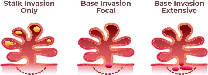
Approaches to substage pT1 disease. TURBT specimens have an inherent lack of orientation which limits precise evaluation of the depth of lamina propria invasion. Even if difficult in some cases, it is recommended to “substage pT1” with the following items: unifocal or multifocal areas of invasion; invasion at the papilla's stalk versus the papillary tumor's base; micrometric evaluation of the largest invasive area (either maximum diameter or depth) with cutoff size of 0.5 mm or 1 mm usually regarded as of prognostic importance


Some authors suggest, and some guidelines cite, the alternative histoanatomic substaging method based on localizing the depth of tumor invasion as superficial or deep (submucosa) using the muscularis mucosae as a reference boundary. It is not practical in daily practice because vesical muscularis mucosae are irregular and discontinuous and may be absent in some bladder regions – such as in the trigone (Paner et al. 2007). Large vessels of lamina propria have been advocated as an alternative reference when there are no muscularis mucosae (Magers et al. 2019). Until new evidence is available, we recommend the stratification of lamina propria invasion into those three parameters listed above (focality, location, and size). Good communication is important in a multidisciplinary team for all those involved to understand the differences in estimating the size of an invasion. Until not defined in guidelines or new staging protocols, using both cutoff sizes of 0.5 mm and 1.0 mm is acceptable. The size of the invasive should be stated as estimated in the largest focus or as an aggregate estimation of all invasive foci. The first is easier to assess and is the base of most studies in the T1 substaging of bladder cancer.
Invasion of muscularis propria is a crucial prognostic information. However the pT2 disease in TURBT specimen indicates the “pT2 stage at a minimum”. The best approach is to comment that this type of specimen is limited to evaluating higher stages.
The invasion of adipose tissue is not equal to pT3 disease in a TURBT specimen. Adipose tissue is a normal finding in lamina propria, between the thick muscle bundles of muscularis propria and in the perivesical tissue (Philip et al. 2000). As a consequence, in most cases the diagnosis of pT3 disease cannot be rendered ina TURBT specimen.
The invasion of the prostatic stroma is not equal to pT4 disease in a TURBT specimen. This observation may reflect the direct invasion of bladder carcinoma into the prostate (true pT4 for a primary bladder tumor). However, urothelial carcinoma is commonly multifocal in bladder mucosa and all urinary tract. Therefore, a TURBT specimen pathologic evaluation cannot distinguish direct invasion from the bladder to prostrate to the scenario of two independent tumors (a bladder primary of any stage, a pT2 urothelial carcinoma of prostatic urethra as primary site). Invasion of prostatic stroma qualifies for pT2 in the case of primary tumor from the prostatic urethra and when there is an extension of an in situ carcinoma of the bladder into the prostatic urethra with the invasion of the prostatic stroma. Invasion of the prostatic stroma may be reported and commented on in light of the observation above (Paner et al. 2017). Correlation with imaging studies should be recommended.
Different noninvasive papillary urothelial carcinoma patterns show peculiar morphology, such as glandular, micropapillary and villoglandular patterns (Amin and Epstein 2012; Miller and Epstein 2009; Lim et al. 2009) (Figs. 14, 15 and 16). Reporting subtypes of noninvasive urothelial carcinoma is optional. However, if reported, it is important to point out in a comment that the biological significance of these findings is unknown and clearly state that it does not refer to subtypes of invasive components (for instance, invasive micropapillary subtype) which infers aggressive behavior.

Noninvasive papillary urothelial carcinoma, glandular pattern. The prognostic value of glandular morphology within the noninvasive component is unknown. This particular case was associated with invasive urothelial carcinoma with glandular differentiation

Noninvasive papillary urothelial carcinoma, micropapillary pattern. The prognostic value of micropapillary morphology within the noninvasive component is unknown

Noninvasive papillary urothelial carcinoma, villoglandular pattern. The prognostic value of villoglandular morphology within the noninvasive component is unknown. Hematoxylin and eosin: 40x magnification (A and C), and 400x magnification (B and D)
Second look TURBT
For samples with residual tumors, all items above apply. As stated, the clinical history of previous diagnoses or treatments is crucial for interpretation. The same is true for whether this is a second look TURB sampling prior to the resection site or a new lesion. PUNLMP diagnosis is restricted for patients with no prior history of urothelial carcinoma (WHO 2022). Urothelial papilloma should be diagnosed with extreme caution in setting a new papillary tumor in the follow-up of urothelial carcinoma. Molecular profiling is recommended before rendering a benign diagnosis in this scenario (WHO 2022). The approach of a recurrent noninvasive tumor after BCG failure depends on the risk of recurrence. Radical cystectomy is recommended for patients with high-risk recurrence (T1 stage, CIS, or high-grade tumors, including some subtypes). Preservation strategies can be offered for patients with high-risk recurrence who are not candidates for or refuse radical cystectomy. Finally, for patients with low-risk recurrence (low-grade Ta), BCG instillation may be offered again, or intravesical chemotherapy (Chang et al. 2016).
The diagnosis of urothelial carcinoma in situ associated with papillary urothelial carcinoma is controversial. WHO 2022 classification states that urothelial carcinoma in situ is rare (only 1–3% are diagnosed de novo). Most cases are diagnosed synchronously or subsequently to high-grade papillary urothelial carcinoma (secondary urothelial carcinoma in situ). However, some authors recommend not diagnosing urothelial carcinoma in situ adjacent to a high-grade papillary urothelial carcinoma (that would be a “shoulder lesion”) or as a residual flat lesion in the previous TURBT site. The International Collaboration on Cancer Reporting (ICCR) reporting guide acknowledges that there are no generally accepted criteria for making this decision but recommends rendering a diagnosis of urothelial carcinoma in situ if: there is a gap of normal urothelium between the papillary tumor and the flat lesion or (b) if the morphology of the flat lesion is different than that of the epithelium on the surface of papillary fronds (Varma et al. 2020).
Such distinction is important for patient care since coexistent urothelial carcinoma in situ will place a high-grade urothelial carcinoma into a high-risk group as per NCCN guideline (NCCN 2024) or very high-risk group as per European Association of Urology guidelines (Lobo et al. 2022). Proper separation of the TURBT site or previous TURBT site biopsy from random biopsies of flat mucosa would easily solve this problem in most cases. It is important, however, to emphasize that the original articles that defined the coexistent of urothelial carcinoma in situ were associated with higher rates of recurrence and progression but did not detail how this coexistence was assessed (adjacent to papillary the tumor vs. away from it) (Sylvester et al. 2006). There is no data on the difference in prognosis of these two different scenarios. Until such information is not available, such distinction will be based on the opinion of experts.
TURBT specimens are usually fragmented and unoriented, which precludes proper evaluation of margins. It is reasonable to infer that the presence of severe atypia in adjacent flat mucosa derives from the extension of papillary carcinoma to the adjacent epithelium. Indeed, concomitant urothelial carcinoma in situ has been shown to have more genetic similarities with high-grade papillary urothelial carcinoma than with urothelial carcinoma in situ signature (Hedegaard et al. 2016). In the case of a specimen clearly referred to as solely a resected tumor, the diagnosis of urothelial carcinoma in situ may indicate that residual disease in non-papillary mucosa is likely.
Although no alternative terminology has been proposed for urothelial carcinoma in situ, many authors understand this lesion as an “extension to flat mucosa of a high-grade papillary urothelial carcinoma.” Such findings should be reported as they may indicate residual disease.
The diagnosis of urothelial carcinoma in situ away from the adjacent mucosa of papillary tumors infers field cancerization of the bladder mucosa, and it is reasonable to understand it as a marker of higher risk of recurrence and progression. Again, the studies that support the coexistence of urothelial carcinoma in situ as a factor in placing a papillary neoplasm in high-risk or very high-risk groups did not clearly separate these two scenarios. Cystoscopic information and separation of samples are crucial for such distinction (see Fig. 1). If this is not the case, the diagnosis of urothelial carcinoma may be rendered with a comment. By analogy, the diagnosis of urothelial carcinoma in situ may be viewed as an intraductal prostate carcinoma. A morphology-defined diagnosis of severe atypia inside prostatic ducts that may either be a precursor lesion or (mostly) the extension of a high-grade carcinoma to pre-existing ducts (Srigley et al. 2022).
In summary, we recommend that the TURBT specimen pathology report should always have an item on the findings of adjacent flat mucosa. Severe atypia in flat mucosa should be diagnosed as urothelial carcinoma. However, ever, interpretation and appropriate comments depend on each situation (see Fig. 1):
-
if the urologist labels the specimen solely as a resection of a papillary tumor, the diagnosis of urothelial carcinoma in situ may be rendered with a comment that it is most probably the extension of a high-grade urothelial carcinoma to adjacent flat bladder mucosa and clinical or prognostic implications are unknown.
-
if the urothelial carcinoma in situ is diagnosed at the site of a previous TURBT (which may be suspected by pathologic evidence of the previous biopsy site and confirmed by clinical information from the urologist), it may be commented that this finding may be a residual (shoulder) lesion of a previous resection of a high-grade papillary urothelial carcinoma.
-
if both a papillary tumor and biopsies of flat mucosa away from the macroscopic tumor were taken and submitted in a single container, the diagnosis of urothelial carcinoma in situ may be rendered with a comment on the limitations to distinguish both situations (and the likely interpretation as shoulder lesion versus field cancerization). ICCR rules may be used and commented on, but the proposed criteria will usually not be met for the diagnosis of urothelial carcinoma in situ.
-
if both a papillary tumor and biopsies of flat mucosa away from the macroscopic tumor were taken and submitted in different containers, the diagnosis of urothelial carcinoma in situ may be rendered straightforward in the flat lesions sent in separately.
Lymphovascular invasion
Lymphovascular invasion (LVI) should be reported in all cases of invasive carcinoma. LVI is an independent predictor of recurrence and decreased overall survival (Streeper et al. 2009). The presence of LVI in TURB specimens has a good concordance with LVI in subsequent cystectomy specimens and is associated with nodal metastasis in nearly 40% of cases (Kunju et al. 2008). The presence of LVI in most cases can be established on morphologic grounds alone. Care must be taken not to overinterpret retraction artifacts as LVI. Superficially invasive urothelial carcinoma commonly displays retraction artifacts around small, irregular nests invading the lamina propria. A similar finding is also seen to a much greater extent in the micropapillary subtype of urothelial carcinoma, in which multiple nests of neoplastic urothelial cells are seen floating inside a space. Therefore, strict morphologic criteria should be used to avoid misinterpretation or retraction artifacts as LVI in the above scenarios. Morphologic criteria were suggested by Algaba in 2006, include tightly cohesive tumors with a smooth border and flattened peripheral cells, completely free-floating / detached tumor thrombus, unequivocal endothelial cell lining, the presence of fibrin and red blood cells around the thrombus, vicinity to an arteriole, and normal surrounding stroma (Algaba 2006).
Flat mucosa (de novo lesion or random biopsies)
Urothelial carcinoma in situ presenting de novo is rare. Most cases are diagnosed in association with papillary urothelial carcinoma, invasive urothelial carcinoma, or in the follow-up of urothelial carcinoma (see discussion above). This diagnosis requires striking nuclear atypia, although it is not required to show the full thickness of urothelium. Urothelial carcinoma in situ is a high-grade lesion and is not graded.
Some flat urothelium may show nuclear atypia and loss of polarity that does not meet the criteria for carcinoma in situ. In these cases, a diagnosis of urothelial dysplasia or flat urothelial atypia of unknown significance may be rendered. Some authors advocate using the term dysplasia only in the setting of follow-up of urothelial carcinoma (Amin et al. 2021). Clinical history is important in this scenario. The progression rate to carcinoma is 14–19% in de novo lesions and 30–36% when diagnosed after a previously resected urothelial carcinoma (Cheng et al. 1999, 2000; Lopez-Beltran et al. 2013).
Some flat urothelium may show no nuclear atypia but architectural changes such as hypercellularity and thickening (more than 6–7 nuclei of polarized urothelial cells), with or without undulation / tented morphology (with no true, thin, and delicate fibrovascular cores). These lesions have been interpreted as a shoulder lesion of low-grade papillary urothelial carcinoma, and in rare instances are observed in a de novo scenario.
The WHO 2022 classification of tumors did not address the classification of such flat lesions. GUPS recommended 2021 the use of the terminology of flat atypical urothelial proliferation and tented atypical urothelial proliferation. It may be interpreted as a potential precursor of noninvasive low-grade papillary urothelial carcinoma or “shoulder”/adjacent/residual lesion of a noninvasive low-grade papillary urothelial carcinoma. Again, clinical history is important for the risk assessment of these lesions. The rate of progression of de novo UPUMP is estimated at 15%. However, the progression rate to carcinoma after a diagnosis of UPUMP may be as high as 41% when there is a prior history of neoplasia. For de novo UPUMP, papillary appearance at cystoscopy and tented (papillary ingrowths) morphology at microscopy were associated with a higher rate of progression (Lowenthal et al. 2020). The use of the terminology urothelial hyperplasia (flat or papillary), UPUMP, and atypical urothelial proliferation (flat or tented) are acceptable, and it is recommended to add a comment on the rates of subsequent low-grade papillary urothelial carcinoma that are influenced by clinical history. Examples of useful comments are available in the template for reporting bladder lesions on the SBP website. Surveillance with cystoscopy is advised.
Additional findings
Non-neoplastic findings are important to be reported for clinicopathologic correlation. Findings may indicate that the procedure reached the previous biopsy site (fibrous scar, foreign body granulomas, eosinophilic cystitis) or that the sample shows effects of previous therapies (different patterns cystitis, caseating granulomas due to infection or previous BCG instillation. Again here, information on previous therapies is important for proper interpretation.
Radiation therapy may induce severe nuclear atypia in urothelial cells. In flat lesions, knowledge of prior radiation exposure and timing is important for the interpretation of the lesion by pathologists. There is a significant overlap between urothelial carcinoma in situ and radiation effect-related changes, and this distinction should not be made within 12 months of radiation therapy (Epstein and Netto 2014). When performing a biopsy in a recently irradiated area, it is important to remove the dystrophic tissue before performing the biopsies.
Some lesions of elevated mucosa or polypoid appearance may be caused by a nephrogenic adenoma, glandular cystitis with or without intestinal metaplasia, or polypoid cystitis. Those lesions must be reported for the urologist to correlate with the findings at cystoscopy.
Squamous metaplasia may be of keratinizing type or no keratinizing type. In the trigone of women, non-keratinizing squamous metaplasia is considered a normal variation of bladder histology. Non-keratinizing squamous metaplasia is usually not associated with the risk of neoplasia or chronic trauma.
Cystectomy specimen
Radical cystectomy specimens may not show residual malignancy in 32–51% of all cases due to previous TURBT resection and/or neoadjuvant chemotherapy (Brimo et al. 2018; Gronostaj et al. 2019). Grossing should be oriented to tumor identification and, if it is not visible, any mucosal or bladder wall abnormality that may indicate a previous biopsy site. If the gross examination is unremarkable, it is recommended to contact the urologist to properly sample or examine the entire region with a previous cancer diagnosis. A randomized sampling of the whole bladder should be made for histological evaluation.
The pathological stage should follow American Joint Committee on Cancer (AJCC) 8th edition (Amin et al. 2017). In the case of no residual invasive tumor, noninvasive papillary urothelial carcinoma (pTa) and urothelial carcinoma in situ (pTis) should be mentioned. Special attention and training in grossing technique are important to identify tumor bed and extravesical mass since the substaging of pT3 is defined grossly.
Treatment effect
Clinical information is absolutely necessary in this situation. A Tumor Regression Grade (TRG) has been proposed and is easy to address: TRG1 (absence of viable cells in a fibrous bed), TRG2 (residual tumor cells occupying < 50% of the tumor bed), and TRG3 (residual tumor cells outgrowing the tumor bed ≥ 50%) (Fleischmann et al. 2014). But the downstage from a muscle invasive tumor to ≤ ypT2 is considered response to neoadjuvant therapy and the grading seems not to affect the prognosis or the patient’s management (Brimo et al. 2018; Gronostaj et al. 2019). Since it is a simple evaluation and shows good reproducibility, we recommend incorporating TRG in pathology reports of cystectomy specimens with known neoadjuvant chemotherapy, with the intention to have data in the future to endorse some studies that have found some prognostic value (Voskuilen et al. 2019; Varma et al. 2020; Paner et al. 2022). It is important to exam multiple sections of the whole thickness of the bladder wall, since it is not rare to see tumor at the perivesical adipose tissue or as a perineural invasion or neoplastic vascular embolus, with no tumor present at the mucosal surface or lamina propria (Fig. 17).

Cystectomy specimen from a patient who underwent neoadjuvant chemotherapy: residual disease only in foci of lymphovascular invasion or at the perivesical adipose tissue. It is important to exam multiple sections of the whole thickness of the bladder wall in a cystectomy specimen from a patient who underwent neoadjuvant chemotherapy. It is not rare to see tumor only in foci of lymphovascular invasion (A) or at the perivesical adipose tissue (B), with no tumor present at the mucosal surface or lamina propria
Additional findings rather than invasive tumors must be reported for clinicopathological correlation. It is important to report urothelial carcinoma in situ since the coexistence of this lesion is a factor in stratifying papillary urothelial carcinomas as high-risk or very high-risk groups. Therefore, urothelial carcinoma in situ with coexistent findings of previous TURBT sites justifies the radical procedure of cystectomy. For clinicopathologic correlation, it is important to report non-neoplastic findings such as those of previous biopsy site (fibrous scar, foreign body granulomas, eosinophilic cystitis) and inflamed flat urothelial mucosa of other sites (different patterns cystitis, caseating granulomas due to infection or previous BCG instillation. Since direct detection of mycobacteria shows poor sensitivity, information on previous BCG therapy is important for proper interpretation to favor a reaction to treatment over true active infection.
Lymph nodes
Lymph nodes may be grossly or microscopically detected in the perivesical fat. The lymph node should be reported as the number of involved lymph nodes and the total number of lymph nodes examined. Grossly positive lymph nodes may be examined with the submission of one section per node. All grossly negative lymph nodes should be entirely submitted, as the presence of the nodal disease may be microscopic and is used as an indication for adjuvant therapy. We follow the ICCR and CAP recommendation to report the size of the largest metastatic deposit, the size of the largest positive lymph node, and the presence/absence of extranodal extension (Grignon et al. 2018; Paner et al. 2022). Some studies have addressed the significance of extranodal extension. Most of these have found the presence of extranodal extension to be associated with disease recurrence or worse cancer-specific survival (Fleischmann et al. 2005; Seiler et al. 2011; Masson-Lecomte et al. 2013; Fajkovic et al. 2013).
Pathological staging requires proper identification of the lymph chains in different containers. Rules of pN classification are pN1 (single regional lymph node metastasis in the true pelvis - perivesical, obturator, internal and external iliac, or sacral lymph node), pN2 (multiple regional lymph node metastases in the true pelvis (same lymph chains, pN3 (metastasis to the common iliac lymph nodes) and pM1a (metastasis limited to lymph nodes beyond the common iliac).
Molecular pathology
From a molecular point of view, bladder carcinomas have two distinct carcinogenesis pathways: the non-invasive carcinoma pathway, also called the papillary pathway, and the invasive urothelial carcinoma pathway or flat pathway. The first is characterized by alterations in genes related to growth factors, transmembrane receptors, and activation of tyrosine kinase pathways such as FGFR, RAS, and PIK3CA-Akt, whereas in the invasive pathway predominates alterations in genes related to the cell cycle, such as p53 (Netto 2011).
Molecular subtypes
Invasive bladder cancer can also be divided into molecular subtypes based on genomic alterations and gene and protein expressions. These molecular subtypes are associated with clinical behavior, histology, and response to treatment.
Several molecular classification systems were proposed. The most well-known molecular classification came from four different Institutions, namely: The Cancer Genome Atlas (TCGA) (Robertson et al. 2017); Lund University (Marzouka et al. 2018); MD Anderson Cancer Center (Choi et al. 2014); and University of North Carolina (Damrauer et al. 2014). Another system that combines these molecular subtypes into a unified system was also proposed by (Kamoun et al. 2020). Generally, these systems’ molecular subtypes are superimposable (Sjödahl et al. 2017). More than 90% of invasive bladder cancers are classified as luminal or basal, although the terminology may differ, particularly within the classification system developed by Lund University. Considering the system developed by TCGA, luminal tumors express high levels of genes associated with urothelial differentiation, such as GATA3 and uroplakines, and low levels of genes associated with basal differentiation, such as high molecular weight keratins and p63. Basal tumors have the opposite expression pattern. The luminal subtype is enriched in tumors with a concomitant non-invasive papillary component and tends to harbor CDKN2A copy number losses and FGFR3 mutations. Basal subtype tumors have a disproportionately high frequency of TP53 mutation. A small subset of invasive bladder cancers lack luminal and basal gene expression and express high levels of neuroendocrine differentiation genes, such as SOX2 and TUBB2B, being referred to as neuronal or small cell, depending on the classification used (Robertson et al. 2017).
FGFR3 gene alterations
FGFR3 alteration is present in about 15% of invasive urothelial carcinomas. Most alterations activate mutations in exon 7 or exon 10, and the minority is composed of rearrangements such as FGRF3::TACC fusion (Robertson et al. 2017).
Mutations in FGFR3 are more common in non-invasive bladder cancer (Sjödahl et al. 2012), particularly non-invasive papillary urothelial carcinoma, corresponding to about 75% of cases (Billerey et al. 2001). In patients with non-invasive bladder cancer, the FGFR3 mutation is associated with better clinical outcomes, specifically with lower progression rates to invasive disease (Van Oers et al. 2007). Mutational status can be heterogeneous in advanced urothelial carcinomas, and it is not uncommon that deep parts of muscle-invasive tumors at TURBT show wild-type FGFR3 while mutations are identified in the superficial component (Pouessel et al. 2016). Therefore, selecting deep parts of the invasive tumor is necessary for evaluating FGFR3 mutations when considering target therapy.
Detecting FGFR3 alteration in advanced urothelial carcinomas, performed by specific PCR test or NGS sequencing, allows targeted treatment with anti-FGFR drugs. Phase II clinical trial results showed objective response rates of approximately 30% in patients with metastasis or unresectable tumor (Loriot et al. 2019).
Current NCCN guidelines (NCCN 2024) recommend considering molecular testing of FGFR3 mutations in stage IIIB bladder cancer (cT1-T4a N2-3) and performing these tests for stage IVA and IVB (metastatic) disease (NCCN 2024). EAU recommends FGFR3 inhibition as an option for second-line therapy or later therapy in patients with proven FGFR3 mutations (strong evidence) (Witjes et al. 2022).
Immuno-oncology and bladder cancer
The ability to evade the immune response is one of the most important characteristics of malignant neoplasms in general. One of the best-known mechanisms of this protection system is through checkpoint inhibitors. Such as the CTLA-4 and PD-1/PD-L1 pathways (Havel et al. 2019). Activation of these systems prevents the cytotoxic response from being effective. Therapeutic agents that block the binding of the inhibitor and its ligand have radically changed the treatment of metastatic bladder cancer, especially in patients who cannot take cisplatin. Several features of bladder cancer are associated with the response to immunotherapy, such as tumor infiltration by CD8 T lymphocytes (Rosenberg et al. 2016) and tumors with high mutational load (Kamat et al. 2017). Some molecular subtypes respond better to immunotherapeutic agents, such as the basal subtype. (Mariathasan et al. 2018). Tumors with genomic instability (Mariathasan et al. 2018) or with tumor infiltration by PD-L1 positive lymphocytes are also more likely to respond to immunotherapy (Balar et al. 2017).
Although no biomarker has a satisfactory predictive capacity, immunohistochemistry’s role in testing for PD-L1 expression has gained prominence to guide the use of these therapies. We have approved several different immunotherapy drugs against urothelial carcinoma by the FDA, each with a particular PD-L1 test. Although PD-L1 expression is not ideal, it can predict immune response and help oncologists make treatment decisions. Oncologists should request the evaluation of PD-L1 expression, always informing which drug they intend to use. Antibody clones and assays and the method of evaluation differ. Depending on which drug will be used, some will require evaluation of PD-L1 expression on inflammatory cells, tumor cells, or both, and the cutoffs are also variable.
Both NCCN and EAU recommend evaluation of PD-L1 expression in cisplatin-ineligible patients. PD-L1 inhibitor atezolizumab is approved for patients with advanced or metastatic UC unfit for cisplatin-based chemotherapy in case of high PD-1 expression defined as tumor-infiltrating immune cells covering > 5% of the tumor area using the SP142 assay. PD-1 inhibitor pembrolizumab is approved for patients with advanced or metastatic UC unfit for any platinum-based chemotherapy in case of high PD-1 expression defined as CPS of > 10 using the Dako 22C33 platform (NCCN 2024; Witjes et al. 2022).
Uro-oncologists may opt to perform early testing of PD-L1 expression and FGFR3 gene alterations to avoid issues of the pre-analytical phase, such as delays in retrieving blocks from different laboratories or testing using old (more than 1-year archived) blocks.
Conclusion
Bladder cancer is an aggressive disease that requires a very meticulous handling in order to identify all factors that will define the tumor behavior. The multidisciplinary involvement is essential for the best result of diagnosis, grading and staging, since the consequences of over or under treatment are immense. In addition, todays personalized medicine requires the adoption of strict analytical protocols based on the best available scientific evidence. This set of recommendations aims to help all specialists to improve communication and patient care.
Availability of data and materials
Not applicable.
Abbreviations
- AJCC:
-
American Joint Committee on Cancer
- CAP:
-
College of American Pathologists
- CD8:
-
Cluster of differentiation 8
- CTLA4:
-
Cytotoxic T-Lymphocyte Associated Protein 4)
- EAU:
-
European Association of Urology
- FDA:
-
(United States) Food and Drug Administration
- ICCR:
-
International Collaboration on Cancer Reporting
- GUPS:
-
Genitourinary Pathology Society
- ISUP:
-
International Society of Urological Pathology
- NCCN:
-
National Comprehensive Cancer Network
- PD-1:
-
Programmed Cell Death (receptor)
- PD-L1:
-
Programmed Cell Death Ligand 1
- PUNLMP:
-
Papillary urothelial neoplasm of low malignant potential
- TURBT:
-
Transurethral resection of a bladder tumor
- UPUMP:
-
Urothelial proliferation of unknown malignant potential
- WHO:
-
World Health Organization
References
Algaba F. Lymphovascular invasion as a prognostic tool for advanced bladder cancer. Curr Opin Urol. 2006;16(5):367–71.
Amin A, Epstein JI. Noninvasive micropapillary urothelial carcinoma: a clinicopathologic study of 18 cases. Hum Pathol. 2012;43:2124–8.
Amin MB, Trpkov K, Lopez-Beltran A, Grignon D, Members of the ISUP Immunohistochemistry in Diagnostic Urologic Pathology Group. Best practices recommendations in the application of immunohistochemistry in the bladder lesions: report from the International Society of Urologic Pathology consensus conference. Am J Surg Pathol. 2014;38:e20-34.
Amin MB, Smith SC, Reuter VE, Epstein JI, Grignon DJ, Hansel DE, Lin O, McKenney JK, Montironi R, Paner GP, Al-Ahmadie HA, Algaba F, Ali S, Alvarado-Cabrero I, Bubendorf L, Cheng L, Cheville JC, Kristiansen G, Cote RJ, Delahunt B, Eble JN, Genega EM, Gulmann C, Hartmann A, Langner C, Lopez-Beltran A, Magi-Galluzzi C, Merce J, Netto GJ, Oliva E, Rao P, Ro JY, Srigley JR, Tickoo SK, Tsuzuki T, Umar SA, Van der Kwast T, Young RH, Soloway MS. Update for the practicing pathologist: the International Consultation on Urologic Disease-European association of urology consultation on bladder cancer. Mod Pathol. 2015;28:612–30.
Amin MB, Edge SB, Greene FL, et al editors. AJCC cancer staging manual. 8th ed. New York: Springer; 2017.
Amin MB, Comperat E, Epstein JI, True LD, Hansel D, Paner GP, Al-Ahmadie H, Baydar D, Bivalacqua T, Brimo F, Cheng L, Cheville J, Dalbagni G, Falzarano S, Gordetsky J, Guo CC, Gupta S, Hes O, Iyer G, Kaushal S, Kunju L, Magi-Galluzzi C, Matoso A, Netto G, Osunkoya AO, Pan CC, Pivovarcikova K, Raspollini MR, Reis H, Rosenberg J, Roupret M, Shah RB, Shariat S, Trpkov K, Weyerer V, Zhou M, McKenney J, Reuter VE. The genitourinary pathology society update on classification and grading of flat and papillary urothelial neoplasia with new reporting recommendations and approach to lesions with mixed and early patterns of Neoplasia. Adv Anat Pathol. 2021;28:179–95.
Assis E. Manual de Boas Práticas em Patologia. São Paulo: Sociedade Brasileira de Patologia; 2020.
Balar AV, Castellano D, O’Donnell PH, Grivas P, Vuky J, Powles T, Plimack ER, Hahn NM, de Wit R, Pang L, Savage MJ, Perini RF, Keefe SM, Bajorin D, Bellmunt J. First-line pembrolizumab in cisplatin-ineligible patients with locally advanced and unresectable or metastatic urothelial cancer (KEYNOTE-052): a multicentre, single-arm, phase 2 study. Lancet Oncol. 2017;18(11):1483–92.
Beltran AL, Cheng L, Montironi R, Blanca A, Leva M, Rouprêt M, Fonseca J, Vidal A, Menendez CL, Pallares J, Bollito E, Reymundo C, Luque RJ, Comperat E. Clinicopathological characteristics and outcome of nested carcinoma of the urinary bladder. Virchows Arch. 2014;465:199–205.
Billerey C, Chopin D, Aubriot-Lorton MH, Ricol D, Gil Diez de Medina S, Van Rhijn B, Bralet MP, Lefrere-Belda MA, Lahaye JB, Abbou CC, Bonaventure J, Zafrani ES, van der Kwast T, Thiery JP, Radvanyi F. Frequent FGFR3 mutations in papillary non-invasive bladder (pTa) tumors. Am J Pathol. 2001;158(6):1955–9.
Bobjer J, Hagberg O, Aljabery F, Gårdmark T, Jahnson S, Jerlström T, Sherif A, Simoulis A, Ströck V, Häggström C, Holmberg L, Liedberg F. Bladder cancer recurrence in papillary urothelial neoplasm of low malignant potential (PUNLMP) compared to G1 WHO 1999: a population-based study. Scand J Urol. 2022;56:14–8.
Brimo F, Downes MR, Jamaspishvili T, Berman D, Barkan GA, Athanazio D, Abro S, Visram K, Yilmaz A, Solanki S, Hahn E, Siemens R, Kassouf W, Trpkov K. Prognostic pathological factors in radical cystectomy after neoadjuvant chemotherapy. Histopathology. 2018;73:732–40.
Chandra A, Griffiths D, McWilliam LJ. Best practice: gross examination and sampling of surgical specimens from the urinary bladder. J Clin Pathol. 2010;63(6):475–9.
Chang SS, Boorjian SA, Chou R, Clark PE, Daneshmand S, Konety BR, Pruthi R, Quale DZ, Ritch CR, Seigne JD, Skinner EC, Smith ND, McKiernan JM. Diagnosis and treatment of non-muscle invasive bladder Cancer: AUA/SUO Guideline. J Urol. 2016;196(4):1021–9.
Cheng L, Cheville JC, Neumann RM, Bostwick DG. Natural history of urothelial dysplasia of the bladder. Am J Surg Pathol. 1999;23:443–7.
Cheng L, Cheville JC, Neumann RM, Bostwick DG. Flat intraepithelial lesions of the urinary bladder. Cancer. 2000;88:625–31.
Choi W, Porten S, Kim S, Willis D, Plimack ER, Hoffman-Censits J, Roth B, Cheng T, Tran M, Lee IL, Melquist J, Bondaruk J, Majewski T, Zhang S, Pretzsch S, Baggerly K, Siefker-Radtke A, Czerniak B, Dinney CP, McConkey DJ. Identification of distinct basal and luminal subtypes of muscle-invasive bladder cancer with different sensitivities to frontline chemotherapy. Cancer Cell. 2014;25(2):152–65.
Colombo R, Hurle R, Moschini M, Freschi M, Colombo P, Colecchia M, Ferrari L, Lucianò R, Conti G, Magnani T, Capogrosso P, Conti A, Pasini L, Burgio G, Guazzoni G, Patriarca C. Feasibility and clinical roles of different substaging systems at first and second transurethral resection in patients with T1 high-grade bladder cancer. Eur Urol Focus. 2018;4(1):87–93.
Damrauer JS, Hoadley KA, Chism DD, Fan C, Tiganelli CJ, Wobker SE, Yeh JJ, Milowsky MI, Iyer G, Parker JS, Kim WY. Intrinsic subtypes of high-grade bladder cancer reflect the hallmarks of breast cancer biology. Proc Natl Acad Sci U S A. 2014;111(8):3110–5.
Epstein JI, Netto GJ. Differential diagnoses in surgical pathology: genitourinary system. Philadelphia: Lippincoty Williams and Wilkins; 2014.
Fajkovic H, Cha EK, Jeldres C, Robinson BD, Rink M, Xylinas E, Chromecki TF, Breinl E, Svatek RS, Donner G, Tagawa ST, Tilki D, Bastian PJ, Karakiewicz PI, Volkmer BG, Novara G, Joual A, Faison T, Sonpavde G, Daneshmand S, Lotan Y, Scherr DS, Shariat SF. Extranodal extension is a powerful prognostic factor in bladder cancer patients with lymph node metastasis. Eur Urol. 2013;64(5):837–45.
Fleischmann A, Thalmann GN, Markwalder R, Studer EU. Extracapsular extension of pelvic lymph node metastases from urothelial carcinoma of the bladder is an independent prognostic factor. J Clin Oncol. 2005;23(10):2358–65.
Fleischmann A, Thalmann GN, Perren A, Seiler R. Tumor regression grade of urothelial bladder cancer after neoadjuvant chemotherapy: a novel and successful strategy to predict survival. Am J Surg Pathol. 2014;38:325–32.
Grignon D, Brimo F, Comperat E, Delahunt B, Koch M, Lopez-Beltran A, Reuter V, Samaratunga H, Shanks J, Tsuzuki T, van der Kwast T, Varma M, Srigley JR. Carcinoma of the bladder, histopathology reporting guide. 1st ed. Australia: International Collaboration on Cancer Reporting; Sydney; 2018.
Gronostaj K, Czech AK, Fronczek J, Drobniak A, Okon K, Chlosta PL, Szczeklik W. The prognostic value of tumor regression grades combined with TNM classification in patients with muscle-invasive bladder cancer who underwent Neoadjuvant chemotherapy followed by radical cystectomy. Clin Genitourin Cancer. 2019;17:e1203-1211.
Hansel DE, Lerner SP. Precision Molecular Pathology of bladder Cancer. In: Cagle PT, editor. Cham: Springer; 2018. https://doi.org/10.1007/978-3-319-64769-2.
Hansel DE, Miller JS, Cookson MS, Chang SS. Challenges in the pathology of non-muscle-invasive bladder cancer: a dialogue between the urologic surgeon and the pathologist. Urology. 2013;81(6):1123–30.
Havel JJ, Chowell D, Chan TA. The evolving landscape of biomarkers for checkpoint inhibitor immunotherapy. Nat Rev Cancer. 2019;19(3):133–50.
Hedegaard J, Lamy P, Nordentoft I, Algaba F, Høyer S, Ulhøi BP, Vang S, Reinert T, Hermann GG, Mogensen K, Thomsen MBH, Nielsen MM, Marquez M, Segersten U, Aine M, Höglund M, Birkenkamp-Demtröder K, Fristrup N, Borre M, Hartmann A, Stöhr R, Wach S, Keck B, Seitz AK, Nawroth R, Maurer T, Tulic C, Simic T, Junker K, Horstmann M, Harving N, Petersen AC, Calle ML, Steyerberg EW, Beukers W, van Kessel KEM, Jensen JB, Pedersen JS, Malmström PU, Malats N, Real FX, Zwarthoff EC, Ørntoft TF, Dyrskjøt L. Comprehensive transcriptional analysis of early-stage urothelial carcinoma. Cancer Cell. 2016;11(1):27–42.
Hentschel AE, van Rhijn BWG, Bründl J, Compérat EM, Plass K, Rodríguez O, Henríquez JDS, Hernández V, de la Peña E, Alemany I, Turturica D, Pisano F, Soria F, Čapoun O, Bauerová L, Pešl M, Bruins HM, Runneboom W, Herdegen S, Breyer J, Brisuda A, Scavarda-Lamberti A, Calatrava A, Rubio-Briones J, Seles M, Mannweiler S, Bosschieter J, Kusuma VRM, Ashabere D, Huebner N, Cotte J, Mertens LS, Cohen D, Lunelli L, Cussenot O, Sheikh SE, Volanis D, Coté JF, Rouprêt M, Haitel A, Shariat SF, Mostafid AH, Nieuwenhuijzen JA, Zigeuner R, Dominguez-Escrig JL, Hacek J, Zlotta AR, Burger M, Evert M, Hulsbergen-van de Kaa CA, van der Heijden AG, Kiemeney LALM, Soukup V, Molinaro L, Gontero P, Llorente C, Algaba F, Palou J, N’Dow J, Babjuk M, van der Kwast TH, Sylvester RJ. Papillary urothelial neoplasm of low malignant potential (PUN-LMP): still a meaningful histo-pathological grade category for Ta, noninvasive bladder tumors in 2019? Urol Oncol. 2020;38:440–8.
Horwich A, Babjuk M, Bellmunt J, Bruins HM, De Reijke TM, De Santis M, Gillessen S, James N, Maclennan S, Palou J, Powles T, Ribal MJ, Shariat SF, Van Der Kwast T, Xylinas E, Agarwal N, Arends T, Bamias A, Birtle A, Black PC, Bochner BH, Bolla M, Boormans JL, Bossi A, Briganti A, Brummelhuis I, Burger M, Castellano D, Cathomas R, Chiti A, Choudhury A, Compérat E, Crabb S, Culine S, De Bari B, DeBlok W, De Visschere PJL, Decaestecker K, Dimitropoulos K, Dominguez-Escrig JL, Fanti S, Fonteyne V, Frydenberg M, Futterer JJ, Gakis G, Geavlete B, Gontero P, Grubmüller B, Hafeez S, Hansel DE, Hartmann A, Hayne D, Henry AM, Hernandez V, Herr H, Herrmann K, Hoskin P, Huguet J, Jereczek-Fossa BA, Jones R, Kamat AM, Khoo V, Kiltie AE, Krege S, Ladoire S, Lara PC, Leliveld A, Linares-Espinós E, Løgager V, Lorch A, Loriot Y, Meijer R, Carmen Mir M, Moschini M, Mostafid H, Müller AC, Müller CR, N’Dow J, Necchi A, Neuzillet Y, Oddens JR, Oldenburg J, Osanto S, Oyen WJG, Pacheco-Figueiredo L, Pappot H, Patel MI, Pieters BR, Plass K, Remzi M, Retz M, Richenberg J, Rink M, Roghmann F, Rosenberg JE, Rouprêt M, Rouvière O, Salembier C, Salminen A, Sargos P, Sengupta S, Sherif A, Smeenk RJ, Smits A, Stenzl A, Thalmann GN, Tombal B, Turkbey B, Vahr Lauridsen S, Valdagni R, Van Der Heijden AG, Van Poppel H, Vartolomei MD, Veskimäe E, Vilaseca A, Vives Rivera FA, Wiegel T, Wiklund P, Williams A, Zigeuner R, Witjes JA. EAU-ESMO consensus statements on the management of advanced and variant bladder cancer-an international collaborative multi-stakeholder effort: under the auspices of the EAU and ESMO guidelines Committees†. Ann Oncol. 2019;30:1697–727.
Kamat AM, Bellmunt J, Galsky MD, Konety BR, Lamm DL, Langham D, Lee CT, Milowsky MI, O’Donnell MA, O’Donnell PH, Petrylak DP, Sharma P, Skinner EC, Sonpavde G, Taylor JA 3rd, Abraham P, Rosenberg JE. Society for Immunotherapy of Cancer consensus statement on immunotherapy for the treatment of bladdercarcinoma. J Immunother Cancer. 2017;5(1):68. https://doi.org/10.1186/s40425-017-0271-0.
Kamoun A, de Reyniès A, Allory Y, Sjödahl G, Robertson AG, Seiler R, Hoadley KA, Groeneveld CS, Al-Ahmadie H, Choi W, Castro MAA, Fontugne J, Eriksson P, Mo Q, Kardos J, Zlotta A, Hartmann A, Dinney CP, Bellmunt J, Powles T, Malats N, Chan KS, Kim WY, McConkey DJ, Black PC, Dyrskjøt L, Höglund M, Lerner SP, Real FX, Radvanyi F. Bladder Cancer Molecular Taxonomy Group. A Consensus Molecular Classification of MuscleinvasiveBladder Cancer. Eur Urol. 2020;77:420–33.
Kunju LP, You L, Zhang Y, Daignault S, Montie JE, Lee CT. Lymphovascular invasion of urothelial cancer in matched transurethral bladder tumor resection and radical cystectomy specimens. J Urol. 2008;180(5):1928–32 (discussion 1932).
Lawless M, Gulati R, Tretiakova M. Stalk versus base invasion in pT1 papillary cancers of the bladder: improved substaging system predicting the risk of progression. Histopathology. 2017;71:406–14.
Leivo MZ, Sahoo D, Hamilton Z, Mirsadraei L, Shabaik A, Parsons JK, Kader AK, Derweesh I, Kane C, Hansel DE. Analysis of T1 bladder cancer on biopsy and transurethral resection specimens: comparison and ranking of T1 quantification approaches to predict progression to muscularis propria invasion. Am J Surg Pathol. 2018;42(1):e1-10.
Lim M, Adsay NV, Grignon D, Osunkoya AO. Urothelial carcinoma with villoglandular differentiation: a study of 14 cases. Mod Pathol. 2009;22(10):1280–6.
Lobo N, Hensley PJ, Bree KK, Nogueras-Gonzalez GM, Navai N, Dinney CP, Sylvester RJ, Kamat AM. Updated European Association of Urology (EAU) prognostic factor risk groups overestimate the risk of progression in patients with non-muscle-invasive bladder Cancer treated with Bacillus Calmette-Guérin. Eur Urol Oncol. 2022;5:84–91.
Lopez-Beltran A, Bassi PF, Pavone-Macaluso M, Montironi R, European Society of Uropathology; Uropathology Working Group. Handling and pathology reporting of specimens with carcinoma of the urinary bladder, ureter, and renal pelvis. A joint proposal of the European Society of Uropathology and the Uropathology Working Group. Virchows Arch. 2004;445(2):103–10.
Lopez-Beltran A, Montironi R, Vidal A, Scarpelli M, Cheng L. Urothelial dysplasia of the bladder: diagnostic features and clinical significance. Anal Quant Cytopathol Histpathol. 2013;35:121–9.
Loriot Y, Necchi A, Park SH, Garcia-Donas J, Huddart R, Burgess E, Fleming M, Rezazadeh A, Mellado B, Varlamov S, Joshi M, Duran I, Tagawa ST, Zakharia Y, Zhong B, Stuyckens K, Santiago-Walker A, De Porre P, O’Hagan A, Avadhani A, Siefker-Radtke AO, BLC2001 Study Group. Erdafitinib in locally advanced or metastatic urothelial carcinoma. N Engl J Med. 2019;381(4):338–48.
Lowenthal BM, Sahoo D, Amin MB, Hansel DE. Urothelial Proliferation of unknown malignant potential involving the bladder: histopathologic features and risk of Progression in De Novo Cases and cases with prior neoplasia. Arch Pathol Lab Med. 2020;144:853–62.
Magers MJ, Lopez-Beltran A, Montironi R, Williamson SR, Kaimakliotis HZ, Cheng L. Staging of bladder cancer. Histopathology. 2019;74:112–34.
Mariathasan S, Turley SJ, Nickles D, Castiglioni A, Yuen K, Wang Y, Kadel EE III, Koeppen H, Astarita JL, Cubas R, Jhunjhunwala S, Banchereau R, Yang Y, Guan Y, Chalouni C, Ziai J, Şenbabaoğlu Y, Santoro S, Sheinson D, Hung J, Giltnane JM, Pierce AA, Mesh K, Lianoglou S, Riegler J, Carano RAD, Eriksson P, Höglund M, Somarriba L, Halligan DL, van der Heijden MS, Loriot Y, Rosenberg JE, Fong L, Mellman I, Chen DS, Green M, Derleth C, Fine GD, Hegde PS, Bourgon R, Powles T. TGFβ attenuates tumour response to PD-L1 blockade by contributing to exclusion of T cells. Nature. 2018;554(7693):544–8.
Marzouka NA, Eriksson P, Rovira C, Liedberg F, Sjödahl G, Höglund M. A validation and extended description of the Lund taxonomy for urothelial carcinoma using the TCGA cohort. Sci Rep. 2018;8(1):3737.
Masson-Lecomte A, Vordos D, Hoznek A, Yiou R, Allory Y, Abbou CC, de la Taille A, Salomon L. External validation of extranodal extension and lymph node density as predictors of survival in node-positive bladder cancer after radical cystectomy. Ann Surg Oncol. 2013;20(4):1389–94.
Maxwell JP, Wang C, Wiebe N, Yilmaz A, Trpkov K. Long-term outcome of primary Papillary Urothelial Neoplasm of low malignant potential (PUNLMP) including PUNLMP with inverted growth. Diagn Pathol. 2015;13:3.
Mazzucchelli R, Marzioni D, Tossetta G, Pepi L, Montironi R. Bladder cancer sample handling and reporting: pathologist’s point of View. Front Surg. 2021;8:754741.
Miller JS, Epstein JI. Noninvasive urothelial carcinoma of the bladder with glandular differentiation: report of 24 cases. Am J Surg Pathol. 2009;33:1241–8.
Miyake M, Nishimura N, Iida K, Fujii T, Nishikawa R, Teraoka S, Takenaka A, Kikuchi H, Abe T, Shinohara N, Okajima E, Shimizu T, Hori S, Tsuchiya N, Owari T, Murakami Y, Taoka R, Kobayashi T, Kojima T, Nishiyama N, Kitamura H, Nishiyama H, Fujimoto K. Intravesical Bacillus Calmette-Guérin treatment for T1 high-Grade non-muscle invasive bladder Cancer with divergent differentiation or variant morphologies. Cancers (Basel). 2021;13:2615.
NCCN Clinical Practice. Guidelines in Oncology (NCCN Guidelines) Guidelines Version 1.2024 Bladder Cancer. 2024.
Netto GJ. Molecular biomarkers in urothelial carcinoma of the bladder: are we there yet? Nat Rev Urol. 2011;9(1):41–51.
Pan CC, Chang YH, Chen KK, Yu HJ, Sun CH, Ho DM. Prognostic significance of the 2004 WHO/ISUP classification for prediction of recurrence, progression, and cancer-specific mortality of non-muscle-invasive urothelial tumors of the urinary bladder: a clinicopathologic study of 1,515 cases. Am J Clin Pathol. 2010;133:788–95.
Paner GP, Ro JY, Wojcik EM, Venkataraman G, Datta MW, Amin MB. Further characterization of the muscle layers and lamina propria of the urinary bladder by systematic histologic mapping: implications for pathologic staging of invasive urothelial carcinoma. Am J Surg Pathol. 2007;31:1420–9.
Paner GP, Montironi R, Amin MB. Challenges in pathologic staging of bladder Cancer: proposals for fresh approaches of assessing pathologic stage in light of recent studies and observations pertaining to bladder histoanatomic variances. Adv Anat Pathol. 2017;24:113–27.
Paner GP, Zhou M, Srigley JR, Harik LR, Amin MB, Allan R, Delahunt B, Bochner BH, Epstein JI, Grignon DJ, Humphrey PA, Montironi R, Pettus J, Reuter VE. Protocol for the Examination of Cystectomy Specimens From Patients With Carcinoma of the Urinary Bladder. College of American Pathologists; 2022.
Philip AT, Amin MB, Tamboli P, Lee TJ, Hill CE, Ro JY. Intravesical adipose tissue: a quantitative study of its presence and location with implications for therapy and prognosis. Am J Surg Pathol. 2000;24(9):1286–90.
Pouessel D, Neuzillet Y, Mertens LS, van der Heijden MS, de Jong J, Sanders J, Peters D, Leroy K, Manceau A, Maille P, Soyeux P, Moktefi A, Semprez F, Vordos D, de la Taille A, Hurst CD, Tomlinson DC, Harnden P, Bostrom PJ, Mirtti T, Horenblas S, Loriot Y, Houédé N, Chevreau C, Beuzeboc P, Shariat SF, Sagalowsky AI, Ashfaq R, Burger M, Jewett MA, Zlotta AR, Broeks A, Bapat B, Knowles MA, Lotan Y, van der Kwast TH, Culine S, Allory Y, van Rhijn BW. Tumor heterogeneity of fibroblast growth factor receptor 3 (FGFR3) mutations in invasive bladder cancer: implications for perioperative anti-FGFR3 treatment. Ann Oncol. 2016;27(7):1311–6.
Reis LO, Taheri D, Chaux A, Guner G, Mendoza Rodriguez MA, Bivalacqua TJ, Schoenberg MP, Epstein JI, Netto GJ. Significance of a minor high-grade component in a low-grade noninvasive papillary urothelial carcinoma of the bladder. Hum Pathol. 2016;47:20–5.
Robertson AG, Kim J, Al-Ahmadie H, Bellmunt J, Guo G, Cherniack AD, Hinoue T, Laird PW, Hoadley KA, Akbani R, Castro MAA, Gibb EA, Kanchi RS, Gordenin DA, Shukla SA, Sanchez-Vega F, Hansel DE, Czerniak BA, Reuter VE, Su X, de Sa Carvalho B, Chagas VS, Mungall KL, Sadeghi S, Pedamallu CS, Lu Y, Klimczak LJ, Zhang J, Choo C, Ojesina AI, Bullman S, Leraas KM, Lichtenberg TM, Wu CJ, Schultz N, Getz G, Meyerson M, Mills GB, McConkey DJ, TCGA Research Network, Weinstein JN, Kwiatkowski DJ, Lerner SP. Comprehensive molecular characterization of muscle-invasive bladder cancer. Cell. 2017;171(3):540-556.e25.
Rosenberg JE, Hoffman-Censits J, Powles T, van der Heijden MS, Balar AV, Necchi A, Dawson N, O’Donnell PH, Balmanoukian A, Loriot Y, Srinivas S, Retz MM, Grivas P, Joseph RW, Galsky MD, Fleming MT, Petrylak DP, Perez-Gracia JL, Burris HA, Castellano D, Canil C, Bellmunt J, Bajorin D, Nickles D, Bourgon R, Frampton GM, Cui N, Mariathasan S, Abidoye O, Fine GD, Dreicer R. Atezolizumab in patients with locally advanced and metastatic urothelial carcinoma who have progressed following treatment with platinum-based chemotherapy: a single-arm, multicentre, phase 2 trial. Lancet. 2016;387(10031):1909–20.
Schultz L. The papilla as a biomarker in the molecular era of bladder oncology. Surg Exp Pathol. 2018;1:10.
Seiler R, von Gunten M, Thalmann GN, Fleischmann A. Extracapsular extension but not the tumour burden of lymph node metastases is an independent adverse risk factor in lymph node-positive bladder cancer. Histopathology. 2011;58(4):571–8.
Sjödahl G, Eriksson P, Liedberg F, Höglund M. Molecular classification of urothelial carcinoma: global mRNA classification versus tumour-cell phenotype classification. J Pathol. 2017;242(1):113–25.
Sjödahl G, Lauss M, Lövgren K, Chebil G, Gudjonsson S, Veerla S, Patschan O, Aine M, Fernö M, Ringnér M, Månsson W, Liedberg F, Lindgren D, Höglund M. A molecular taxonomy for urothelial carcinoma. Clin Cancer Res. 2012;18(12):3377–86.
Srigley JR, Tsuzuki T, Amin MB, Rubin MA, editors. Intraductal carcinoma of the prostate. In: WHO Classification of Tumours Editorial Board. Urinary and male genital tumours. Lyon (France): International Agency for Research on Cancer; 2022. (WHO classification of tumours series, 5th ed.; vol. 8). https://publications.iarc.fr .
Streeper NM, Simons CM, Konety BR, Muirhead DM, Williams RD, O’Donnell MA, Joudi FN. The significance of lymphovascular invasion in transurethral resection of bladder tumor and cystectomy specimens on the survival of patients with urothelial bladder cancer. BJU Int. 2009;103(4):475–9.
Sylvester RJ, van der Meijden AP, Oosterlinck W, Witjes JA, Bouffioux C, Denis L, Newling DW, Kurth K. Predicting recurrence and progression in individual patients with stage Ta T1 bladder cancer using EORTC risk tables: a combined analysis of 2596 patients from seven EORTC trials. Eur Urol. 2006;49:466–465 discussion 475-7.
Toll AD, Epstein JI. Invasive low-grade papillary urothelial carcinoma: a clinicopathologic analysis of 41 cases. Am J Surg Pathol. 2012;36:1081–6.
van de Fransen EE, Otto W, Hartmann A, Bertz S, Mayr R, Bründl J, Breyer J, Manach Q, Compérat EM, Boormans JL, Bosschieter J, Jewett MAS, Stoehr R, van Leenders GJLH, Nieuwenhuijzen JA, Zlotta AR, Hendricksen K, Rouprêt M, Burger M, van der Kwast TH, van Rhijn BWG. Metric substage according to micro and extensive lamina propria invasion improves prognostics in T1 bladder cancer. Urol Oncol. 2018;36(8):361.e7-361.e13.
van Oers JM, Wild PJ, Burger M, Denzinger S, Stoehr R, Rosskopf E, Hofstaedter F, Steyerberg EW, Klinkhammer-Schalke M, Zwarthoff EC, van der Kwast TH, Hartmann A. FGFR3 mutations and a normal CK20 staining pattern define low-grade noninvasive urothelial bladder tumours. Eur Urol. 2007;52(3):760–8.
Van Rhijn BW, van der Kwast TH, Alkhateeb SS, Fleshner NE, van Leenders GJ, Bostrom PJ, van der Aa MN, Kakiashvili DM, Bangma CH, Jewett MA, Zlotta AR. A new and highly prognostic system to discern T1 bladder cancer substage. Eur Urol. 2012;61(2):378–84.
Varma M, Srigley JR, Brimo F, Compérat E, Delahunt B, Koch M, Lopez-Beltran A, Reuter V, Samaratunga H, Shanks JH, Tsuzuki T, van der Kwast T, Webster F, Grignon D. Dataset for the reporting of urinary tract carcinoma-biopsy and transurethral resection specimen: recommendations from the International collaboration on Cancer Reporting (ICCR). Mod Pathol. 2020;33:700–12.
Voskuilen CS, Oo HZ, Genitsch V, Smit LA, Vidal A, Meneses M, Necchi A, Colecchia M, Xylinas E, Fontugne J, Sibony M, Rouprêt M, Lenfant L, Côté JF, Buser L, Saba K, Furrer MA, van der Heijden MS, Daugaard M, Black PC, van Rhijn BWG, Hendricksen K, Poyet C, Seiler R. Multicenter Validation of histopathologic tumor regression grade after neoadjuvant chemotherapy in muscle-invasive bladder carcinoma. Am J Surg Pathol. 2019;43:1600–10.
Warrick JI, Knowles MA, Yves A, van der Kwast T, Grignon DJ, Kristiansen G, Egevad L, Hartmann A, Cheng L. Report from the International Society of Urological Pathology (ISUP) consultation conference on molecular pathology of urogenital cancers. II. Molecular pathology of bladder cancer: progress and challenges. Am J Surg Pathol. 2020;44:e30–46.
Wasco MJ, Daignault S, Bradley D, Shah RB. Nested variant of urothelial carcinoma: a clinicopathologic and immunohistochemical study of 30 pure and mixed cases. Hum Pathol. 2010;41:163–71.
WHO Classification of Tumours Editorial Board. Urinary and male genital tumors [Internet]. Lyon (France): International Agency for Research on Cancer; 2022 (WHO classification of tumors series, 5th ed.; vol. 8). https://tumourclassification.iarc.who.int/chapters/36.
Witjes JA, Bruins HM, Carrión A, Cathomas R, Compérat EM, Efstathiou JA, Fietkau R, Gakis G, van der Heijden AG, Lorch A, Meijer RP, Milowsky MI, Panebianco V, Rink M, Thalmann GN, Veskimäe E. EAU Guidelines on Muscle-invasive and Metastatic Bladder Cancer. 2022.
Acknowledgements
None.
Funding
This study was funded by the Brazilian Society of Pathology, which funded six consensus meetings.
Author information
Authors and Affiliations
Contributions
EA and ICS conceived the idea. DAA, LSA, and IWC were significant contributors to the writing of the manuscript. All authors were major contributors to critically revising the manuscript for important intellectual content. All authors read and approved the final manuscript.
Corresponding author
Ethics declarations
Ethics approval and consent to participate
Not applicable.
Consent for publication
Not applicable.
Competing interests
The authors declare that they have no competing interests.
Additional information
Publisher’s Note
Springer Nature remains neutral with regard to jurisdictional claims in published maps and institutional affiliations.
Supplementary Information
Rights and permissions
Open Access This article is licensed under a Creative Commons Attribution 4.0 International License, which permits use, sharing, adaptation, distribution and reproduction in any medium or format, as long as you give appropriate credit to the original author(s) and the source, provide a link to the Creative Commons licence, and indicate if changes were made. The images or other third party material in this article are included in the article's Creative Commons licence, unless indicated otherwise in a credit line to the material. If material is not included in the article's Creative Commons licence and your intended use is not permitted by statutory regulation or exceeds the permitted use, you will need to obtain permission directly from the copyright holder. To view a copy of this licence, visit http://creativecommons.org/licenses/by/4.0/.
About this article

Cite this article
Athanazio, D.A., Amorim, L.S., da Cunha, I.W. et al. Handling and pathology reporting guidelines for bladder epithelial neoplasms – recommendations from the Brazilian Society of Pathology / Brazilian Society of Urology / Brazilian Society of Clinical Oncology. Surg Exp Pathol 7, 8 (2024). https://doi.org/10.1186/s42047-024-00151-7
Received:
Accepted:
Published:
DOI: https://doi.org/10.1186/s42047-024-00151-7