- Research
- Open access
- Published:
Bubbles in the belly- a case based approach to cystic peritoneal masses
Surgical and Experimental Pathology volume 4, Article number: 17 (2021)
Abstract
Background
Primary cystic neoplasms of the peritoneum are rare lesions and not commonly encountered in practice. Many intra-abdominal processes may mimic cystic masses within the peritoneal cavity and pose a diagnostic challenge to both the pathologist and radiologist. Clinical presentation is diverse and varied. These lesions are usually benign. Hence complete surgical excision is the treatment of choice in most of the cases.
Methods
Study design: Descriptive Retrospective study.
Cystic peritoneal lesions were identified and studied from data over a period of 5 years in the Histopathology Section at a tertiary care hospital in Pune, India. Mode of presentation, imaging findings in addition to gross and histopathologic findings of these lesions were studied.
Results
Out of 50 peritoneal lesions studied over a period of 5 years, only 7 were identified to be cystic peritoneal masses.
Of these two were found to be peritoneal cysts, two mesenteric cysts, one an infected mesenteric cyst and one each a mucinous cystadenoma and lymphangioma.
Conclusions
Correct diagnosis rests in the hands of the pathologist and ensures that the patient receives appropriate and timely management. Hence knowledge of the spectrum of these rare cystic peritoneal masses is necessary to distinguish from other potential cystic abdominal mimicker masses and avoid a potential pitfall.
Introduction
Primary cystic neoplasms of the peritoneum are uncommon lesions, rarely encountered in daily practice. (Liew et al., 1994) Most of these lesions present with vague complaints of abdominal pain and nausea. Less commonly they may present with bowel obstruction due to external compression of the bowel. (Mackenzie et al., 1993) Because of variable and non-specific clinical symptoms and signs, many of them are discovered either accidentally during an abdominal radiological examination performed for some other reason or during laparotomy for the management of one of the complications. (Mishra et al., 2018) There are many intra-abdominal processes which may mimic cystic masses within the peritoneal cavity. These include the various fluid collections such as abscesses, seroma, biloma, urinoma, or lymphoceles. These are often recognized based on relevant clinical history like recent surgery or trauma. (Arraiza et al., 2015) Cystic peritoneal masses may be localized anywhere in the mesentery, from duodenum to rectum, however, these are mostly found in the ileum and right colon mesentery. (Huis et al., 2002) Complete surgical excision of the cyst is usually the treatment of choice, with no recurrence post excision. (Pithawa et al., 2014) Hence knowledge of the spectrum of these rare cystic peritoneal masses is necessary to distinguish from other potential cystic abdominal mimicker masses and avoid a potential pitfall. (6).
Potential problems include misdiagnosis at imaging, as well as confusion over organ of origin- mainly pancreas and ovaries. A careful gross and histopathologic approach helps to overcome this.
Aims and objectives
-
1)
To study the varied presentation of cystic peritoneal masses.
-
2)
To study the gross and histopathological findings of cystic peritoneal masses.
Materials and methods
Study design: descriptive retrospective study
Cystic peritoneal lesions were identified and studied from data over a period of 5 years, from 2016 to 2020 in the Histopathology Section at a tertiary care hospital in Pune, India.
Inclusion Criteria: All surgically excised cystic lesions of the peritoneum were included in our study.
Exclusion criteria: Cystic neoplasms of the pancreas, ovary and organs other than peritoneum were excluded. Mode of presentation, imaging findings in addition to gross and histopathologic findings of these lesions were studied.
Results
Out of 50 peritoneal lesions studied over a period of 5 years, only 7 were identified to be cystic peritoneal masses.
Of these two were found to be peritoneal cysts, two mesenteric cysts, one an infected mesenteric cyst and one each a mucinous cystadenoma and lymphangioma.
Cystic peritoneal masses were more commonly found in female (5/7) and were more commonly seen in females of reproductive age group. Two lesions were seen in the paediatric age.
Most lesions were associated with symptoms at presentation in the form of either abdominal pain or abdominal fullness while two were discovered incidentally at surgery.
On gross examination 5 out of 7 lesions were larger than 10 cm in diameter while only one was 1 cm in diameter.
Most of the lesions were excised and received intact.
Peritoneal cysts tended to be intact on cut opening while mesenteric cysts, mucinous cystadenoma and lymphangiomas tended to be multicystic multiloculated.
All the lesions had smooth inner surfaces without any solid areas or papillary excrescence signifying that they were all benign in nature.
Of the 7 lesions identified only 1 required immunohistochemistry for confirmation of diagnosis.
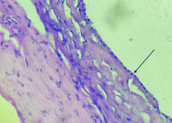
Immunohistochemistry was resorted to in only one case, panel used was CD34 and Calretinin (Table 1 and Figs. 1, 2, 3, 4, 5, 6, 7, 8, 9 and 10).
Discussion
Cystic peritoneal masses have been classified based on their lining on histology into four categories—endothelial, epithelial, mesothelial, and others (germ cell tumours, sex cord gonadal stromal tumours, cystic mesenchymal tumours, fibrous wall tumours, and infectious cystic peritoneal lesions). (8).
They arise from the mesothelial lining of the peritoneum.
Peritoneal inclusion cysts are the most common among cystic peritoneal neoplasms. They were seen in five of our cases.
These are usually discovered incidentally and are solitary unilocular, thinned walled and contain serous fluid. Two of our cases were asymptomatic at presentation and the cysts were discovered incidentally in both patients who had undergone a lower segment Caesarean section. The remaining cases complained of either abdominal distension or abdominal pain. Five of the six cases were reproductive age females while one was an elderly male. Most of the cysts were large on gross examination measuring more than10 cm in size. Histologically these are lined by simple flattened epithelium resting on a fibrocollagenous stroma. (Sudiono et al., 2016) We had a case on infected mesenteric cyst present to us. This complication is commonly encountered in reproductive age women. In our histologically there was focal to dense inflammatory infiltrate along with giant cell reaction. In doubtful cases IHC can be resorted to. The lining cells are positive for calretinin, WT1 and D2–40, and negative for Pax8 and Ber-Ep4.
Lymphangiomas are congenital benign vascular lesions resulting from developmental failure of the lymphatic system. They occur more commonly in children though adult clinical presentation is not uncommon (Levy et al. 2004). The pathogenesis is vague but may represent embryologic remnants of lymphatic tissues with aberrant or obstructed outflow or arise from lymph sacs sequestered during development (Park et al. 1999). From a study of 14 patients (Goh et al. 2005) hypothesized that endogenous estrogens might play a role in the enlargement or growth of lymphangiomas, thereby explaining the female preponderance of intraabdominal lymphangiomas in adults. Lymphangiomatosis is a rare disease with multifocal sites of lymphatic proliferation that typically presents during childhood and may involve multiple parenchymal organs including the lung, liver, spleen, bone, and skin. Histologically, lymphangiomas are thin-walled cystic masses with a smooth grey, pink, tan, or yellow external surface. On cut section, they may contain large macroscopic interconnecting cysts (often referred to as cystic hygroma or cystic lymphangioma or microscopic cysts (cavernous lymphangioma) (Sarno et al. 1984). The cysts may contain chylous, serous, haemorrhagic, or mixed fluid (Ros et al. 1987). The dilated lymphatic spaces are lined with endothelial cells resembling the cells that line normal lymphatics. The supporting stroma, about 70% of lymphangiomas occur in the head and neck; 20% in axillary regions; and the remaining 5% are in the mesentery, retroperitoneum, abdominal viscera, lung, and mediastinum (Sarno et al. 1984). Abdominal lymphangiomas occur most commonly in the mesentery, followed by the omentum, mesocolon, and retroperitoneum (Levy et al. 2004). We had one case of intraperitoneal lymphangioma reported. The patient was an 8-year-old male who complained on abdominal pain. Imaging showed a large well defined, thin-walled cystic mass lesion with thin internal enhancing septae. An intact 17x14x10 cm tense globular cyst was received for histopathological examination. On cut section the cyst was bilocular with smooth inner surface and brownish fluid. Microscopy showed dilated lymphatic channels alongwith lymphoid aggregates. Immunohistochemistry was resorted to and showded CD 34 positivity and calretinin negative. This confirmed the diagnosis of Lymphangioma.
Histologically lymphangiomas are characterised by large lymphatic channels in loose connective tissue stroma, focally disorganized smooth muscle in wall of larger channels and peripheral lymphoid aggregates.
Immunohistochemically these are positive for CD 31, CD 34, Desmin and negative for Calretinin, WT- 1 and Cytokeratinin 5/6 (Nagata et al., 2014).
Primary retroperitoneal and intraperitoneal cystadenomas are the rarest, even though they are common in the ovaries Their histogenesis is uncertain and several theories have been proposed, with most authors suggesting that they develop through mucinous metaplasia in a pre-existing mesothelium-lined cyst. (Singh et al., 2015) These can be commonly confused for ovarian mucinous cystadenomas. We had one case of intraperitoneal mucinous cystadenoma in a 28-year-old female. She complained of epigastric pain for 2 months. Classically the pain increased on sitting and was relieved on lying down. No history of trauma was elicited.
She had a history of pancreatic cyst about 7 years ago for which a cystogastrostomy was done. Imaging was suggestive of either mesenteric cyst or lymphangioma as possible differentials. We had received a an intact 18x14x12 cm, encapsulated mass, which on cut section was multicystic and multiloculated. Mucinous fluid oozes out on cut section and no solid areas were identified. Histopathology showed a Mucinous Cystadenoma showing a cyst wall with lining epithelium composed of benign looking goblet cells with collagenised stroma.
Intraperitoneal mucinous cystadenomas exhibit benign behaviour and surgical excision is usually curative. Grossly these are well encapsulated, bosselated and multiloculated. On cut opening they show mucinous material. Histologically these are lined by goblet cells in a picket fence pattern that rest on a fibrocollagenised stroma. Sometimes focal atypia may be noted and there may be evidence of ovarian stroma in all. Our case did not exhibit cytologic atypia. Immunohistochemically there is positivity for Carcinoembryonic Antigen, Epithelial Membrane Antigen and ck 7+/ck 20 -.
On follow up visits none of our cases reported any recurrence.
Take home message
Cystic peritoneal masses are exceedingly rare and are commonly confused for ovarian neoplasms, pancreatic neoplasms, etc. both clinically and radiologically. Clinically they may be discovered incidentally at surgery or may present rarely as acute abdomen. Many of these lesions are commonly encountered in woman of reproductive age groups and are seen to commonly arise in relation to the pelvis and hence commonly confused with ovarian neoplasms. They can be confused with pancreatic neoplasms as well. These could serve as a potential pitfall and be misdiagnosed both during imaging and clinically. Most of the cystic peritoneal masses are clinically benign and do not recur post-surgical excision.
Under such situations the final diagnosis rests on the histopathological analysis.
IHC should be resorted to in doubtful cases.
Hence it is essential that pathologists familiarise themselves with the classification and potential differential diagnoses of cystic peritoneal masses to guide appropriate clinical care.
This would also save the patients from undergoing additional investigations and ‘cancer scare’ particularly in relation to pancreatic and ovarian neoplasms that tend to be more aggressive in behavior.
Conclusion
Cystic Peritoneal Masses are uncommon neoplasms and can easily be misdiagnosed both clinically and during imaging. The correct diagnoses rests in the hands of the surgical pathologist. These are mostly benign neoplasms and complete surgical excision is curative. Hence it is essential that they are diagnosed correctly for appropriate management.
Availability of data and materials
Data sharing is not applicable as no data sets were analysed or generated during this study.
Abbreviations
- IHC:
-
Immunohistochemistry
- CT:
-
Computed tomography
- HPE:
-
Histopathological examination
- USG:
-
Ultrasound
- L.S.C.S.:
-
Lower Segment Caeserean Section
- LP:
-
Lower power (100x magnification)
- HP:
-
High power (400x magnification)
References
Arraiza M, Metser U, Vajpeyi R, Khalili K, Hanbidge A, Kennedy E, Ghai S. Primary cystic peritoneal masses and mimickers: spectrum of diseases with pathologic correlation. Abdom Imaging. 2015;40(4):875–906. https://doi.org/10.1007/s00261-014-0250-6 PMID: 25269999.
Huis M, Balija M, Lez C, Szerda F, Stulhofer M. Ciste mezenterija [mesenteric cysts]. Acta Med Croatica. 2002;56(3):119–24 Croatian. PMID: 12630343.
Liew SC, Glenn DC, Storey DW. Mesenteric cyst. Aust N Z J Surg. 1994;64(11):741–4. https://doi.org/10.1111/j.1445-2197.1994.tb04530.x PMID: 7945079.
Mackenzie DJ, Shapiro SJ, Gordon LA, Ress R. Laparoscopic excision of a mesenteric cyst. J Laparoendosc Surg. 1993;3(3):295–9. 8347889. https://doi.org/10.1089/lps.1993.3.295.
Mishra T, Karegar MM, Rojekar AV, Joshi AS. Multilocular peritoneal inclusion cyst, rare occurrence in men: a case report. Indian J Pathol Microbiol. 2018;61(1):164–6. https://doi.org/10.4103/IJPM.IJPM_480_16.
Nagata H, Yonemura Y, Canbay E, Ishibashi H, Narita M, Mike M, Kano N. Differentiating a large abdominal cystic lymphangioma from multicystic mesothelioma: report of a case. Surg Today. 2014;44(7):1367–70. https://doi.org/10.1007/s00595-013-0654-x Epub 2013 Jun 27. PMID: 23807639.
Pithawa AK, Bansal AS, Kochar SP. Mesenteric cyst: A rare intra-abdominal tumour. Med J Armed Forces India. 2014;70(1):79–82. https://doi.org/10.1016/j.mjafi.2012.06.010 Epub 2012 Oct 23. PMID: 24936122; PMCID: PMC4054796.
Singh A, Sehgal A, Mohan H. Multilocular peritoneal inclusion cyst mimicking an ovarian tumor: A case report. J Midlife Health. 2015;6(1):39–40. https://doi.org/10.4103/0976-7800.153648 PMID: 25861208; PMCID: PMC4389384.
Sudiono DR, Ponten JB, Zijta FM. Acute abdominal pain caused by an infected mesenteric cyst in a 24-year-old female. Case Rep Radiol. 2016;2016:4. https://doi.org/10.1155/2016/8437832.
Acknowledgements
Not applicable.
Funding
Not applicable.
Author information
Authors and Affiliations
Contributions
Dr. Savita S. Patil – First author, reporting of cases and review of draft. Dr. Parth B. Shah – Second and Corresponding author, data collection, data compilation and analysis, preparation, write-up and drafting of manuscript. Dr. Jyoti K. Kudrimoti- Third author- Review of draft. Dr. Leena A. Nakate- Fourth author- Review of draft. The authors read and approved the final manuscript.
Corresponding author
Ethics declarations
Ethics approval and consent to participate
Not applicable.
Data was analysed retrospectively and patient information has been kept confidential,
Consent for publication
Not applicable.
Competing interests
The authors declare that they have no competing interests.
Additional information
Publisher’s Note
Springer Nature remains neutral with regard to jurisdictional claims in published maps and institutional affiliations.
Rights and permissions
Open Access This article is licensed under a Creative Commons Attribution 4.0 International License, which permits use, sharing, adaptation, distribution and reproduction in any medium or format, as long as you give appropriate credit to the original author(s) and the source, provide a link to the Creative Commons licence, and indicate if changes were made. The images or other third party material in this article are included in the article's Creative Commons licence, unless indicated otherwise in a credit line to the material. If material is not included in the article's Creative Commons licence and your intended use is not permitted by statutory regulation or exceeds the permitted use, you will need to obtain permission directly from the copyright holder. To view a copy of this licence, visit http://creativecommons.org/licenses/by/4.0/.
About this article
Cite this article
Patil, S.S., Shah, P.B., Kudrimoti, J.K. et al. Bubbles in the belly- a case based approach to cystic peritoneal masses. Surg Exp Pathol 4, 17 (2021). https://doi.org/10.1186/s42047-021-00099-y
Received:
Accepted:
Published:
DOI: https://doi.org/10.1186/s42047-021-00099-y


